This old version of Proteopedia is provided for student assignments while the new version is undergoing repairs. Content and edits done in this old version of Proteopedia after March 1, 2026 will eventually be lost when it is retired in about June of 2026.
Apply for new accounts at the new Proteopedia. Your logins will work in both the old and new versions.
3ouh
From Proteopedia
(Difference between revisions)
(New page: 200px <!-- The line below this paragraph, containing "STRUCTURE_3ouh", creates the "Structure Box" on the page. You may change the PDB parameter (which sets the PD...) |
|||
| (7 intermediate revisions not shown.) | |||
| Line 1: | Line 1: | ||
| - | [[Image:3ouh.png|left|200px]] | ||
| - | + | ==PHD2-R127 with JNJ41536014== | |
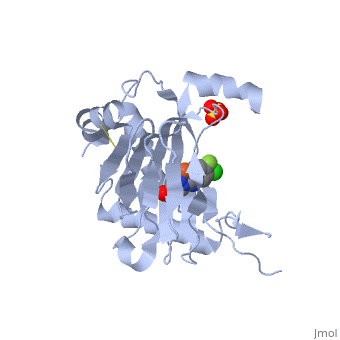
| - | + | <StructureSection load='3ouh' size='340' side='right'caption='[[3ouh]], [[Resolution|resolution]] 2.51Å' scene=''> | |
| - | + | == Structural highlights == | |
| - | + | <table><tr><td colspan='2'>[[3ouh]] is a 1 chain structure with sequence from [https://en.wikipedia.org/wiki/Homo_sapiens Homo sapiens]. Full crystallographic information is available from [http://oca.weizmann.ac.il/oca-bin/ocashort?id=3OUH OCA]. For a <b>guided tour on the structure components</b> use [https://proteopedia.org/fgij/fg.htm?mol=3OUH FirstGlance]. <br> | |
| - | + | </td></tr><tr id='method'><td class="sblockLbl"><b>[[Empirical_models|Method:]]</b></td><td class="sblockDat" id="methodDat">X-ray diffraction, [[Resolution|Resolution]] 2.51Å</td></tr> | |
| - | + | <tr id='ligand'><td class="sblockLbl"><b>[[Ligand|Ligands:]]</b></td><td class="sblockDat" id="ligandDat"><scene name='pdbligand=014:1-(5-CHLORO-6-FLUORO-1H-BENZIMIDAZOL-2-YL)-1H-PYRAZOLE-4-CARBOXYLIC+ACID'>014</scene>, <scene name='pdbligand=FE2:FE+(II)+ION'>FE2</scene>, <scene name='pdbligand=SO4:SULFATE+ION'>SO4</scene></td></tr> | |
| - | + | <tr id='resources'><td class="sblockLbl"><b>Resources:</b></td><td class="sblockDat"><span class='plainlinks'>[https://proteopedia.org/fgij/fg.htm?mol=3ouh FirstGlance], [http://oca.weizmann.ac.il/oca-bin/ocaids?id=3ouh OCA], [https://pdbe.org/3ouh PDBe], [https://www.rcsb.org/pdb/explore.do?structureId=3ouh RCSB], [https://www.ebi.ac.uk/pdbsum/3ouh PDBsum], [https://prosat.h-its.org/prosat/prosatexe?pdbcode=3ouh ProSAT]</span></td></tr> | |
| - | + | </table> | |
| - | + | == Disease == | |
| - | + | [https://www.uniprot.org/uniprot/EGLN1_HUMAN EGLN1_HUMAN] Defects in EGLN1 are the cause of familial erythrocytosis type 3 (ECYT3) [MIM:[https://omim.org/entry/609820 609820]. ECYT3 is an autosomal dominant disorder characterized by increased serum red blood cell mass, elevated serum hemoglobin and hematocrit, and normal serum erythropoietin levels.<ref>PMID:16407130</ref> <ref>PMID:17579185</ref> | |
| - | + | == Function == | |
| - | == | + | [https://www.uniprot.org/uniprot/EGLN1_HUMAN EGLN1_HUMAN] Cellular oxygen sensor that catalyzes, under normoxic conditions, the post-translational formation of 4-hydroxyproline in hypoxia-inducible factor (HIF) alpha proteins. Hydroxylates a specific proline found in each of the oxygen-dependent degradation (ODD) domains (N-terminal, NODD, and C-terminal, CODD) of HIF1A. Also hydroxylates HIF2A. Has a preference for the CODD site for both HIF1A and HIF1B. Hydroxylated HIFs are then targeted for proteasomal degradation via the von Hippel-Lindau ubiquitination complex. Under hypoxic conditions, the hydroxylation reaction is attenuated allowing HIFs to escape degradation resulting in their translocation to the nucleus, heterodimerization with HIF1B, and increased expression of hypoxy-inducible genes. EGLN1 is the most important isozyme under normoxia and, through regulating the stability of HIF1, involved in various hypoxia-influenced processes such as angiogenesis in retinal and cardiac functionality.<ref>PMID:11595184</ref> <ref>PMID:12351678</ref> <ref>PMID:15897452</ref> <ref>PMID:19339211</ref> <ref>PMID:21792862</ref> |
| - | [[3ouh]] | + | |
==See Also== | ==See Also== | ||
| - | *[[ | + | *[[Polyl hydroxylase domain 3D structures|Polyl hydroxylase domain 3D structures]] |
*[[Prolyl hydroxylase domain|Prolyl hydroxylase domain]] | *[[Prolyl hydroxylase domain|Prolyl hydroxylase domain]] | ||
| + | == References == | ||
| + | <references/> | ||
| + | __TOC__ | ||
| + | </StructureSection> | ||
[[Category: Homo sapiens]] | [[Category: Homo sapiens]] | ||
| - | [[Category: | + | [[Category: Large Structures]] |
| - | [[Category: | + | [[Category: Clark R]] |
| - | [[Category: | + | [[Category: Kim H]] |
| - | + | ||
Current revision
PHD2-R127 with JNJ41536014
| |||||||||||
