This old version of Proteopedia is provided for student assignments while the new version is undergoing repairs. Content and edits done in this old version of Proteopedia after March 1, 2026 will eventually be lost when it is retired in about June of 2026.
Apply for new accounts at the new Proteopedia. Your logins will work in both the old and new versions.
1lvl
From Proteopedia
(Difference between revisions)
(New page: 200px<br /><applet load="1lvl" size="450" color="white" frame="true" align="right" spinBox="true" caption="1lvl, resolution 2.45Å" /> '''THE REFINED STRUCTUR...) |
|||
| (17 intermediate revisions not shown.) | |||
| Line 1: | Line 1: | ||
| - | [[Image:1lvl.gif|left|200px]]<br /><applet load="1lvl" size="450" color="white" frame="true" align="right" spinBox="true" | ||
| - | caption="1lvl, resolution 2.45Å" /> | ||
| - | '''THE REFINED STRUCTURE OF PSEUDOMONAS PUTIDA LIPOAMIDE DEHYDROGENASE COMPLEXED WITH NAD+ AT 2.45 ANGSTROMS RESOLUTION'''<br /> | ||
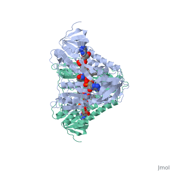
| - | == | + | ==THE REFINED STRUCTURE OF PSEUDOMONAS PUTIDA LIPOAMIDE DEHYDROGENASE COMPLEXED WITH NAD+ AT 2.45 ANGSTROMS RESOLUTION== |
| - | + | <StructureSection load='1lvl' size='340' side='right'caption='[[1lvl]], [[Resolution|resolution]] 2.45Å' scene=''> | |
| + | == Structural highlights == | ||
| + | <table><tr><td colspan='2'>[[1lvl]] is a 1 chain structure with sequence from [https://en.wikipedia.org/wiki/Pseudomonas_putida Pseudomonas putida]. Full crystallographic information is available from [http://oca.weizmann.ac.il/oca-bin/ocashort?id=1LVL OCA]. For a <b>guided tour on the structure components</b> use [https://proteopedia.org/fgij/fg.htm?mol=1LVL FirstGlance]. <br> | ||
| + | </td></tr><tr id='method'><td class="sblockLbl"><b>[[Empirical_models|Method:]]</b></td><td class="sblockDat" id="methodDat">X-ray diffraction, [[Resolution|Resolution]] 2.45Å</td></tr> | ||
| + | <tr id='ligand'><td class="sblockLbl"><b>[[Ligand|Ligands:]]</b></td><td class="sblockDat" id="ligandDat"><scene name='pdbligand=FAD:FLAVIN-ADENINE+DINUCLEOTIDE'>FAD</scene>, <scene name='pdbligand=NAD:NICOTINAMIDE-ADENINE-DINUCLEOTIDE'>NAD</scene></td></tr> | ||
| + | <tr id='resources'><td class="sblockLbl"><b>Resources:</b></td><td class="sblockDat"><span class='plainlinks'>[https://proteopedia.org/fgij/fg.htm?mol=1lvl FirstGlance], [http://oca.weizmann.ac.il/oca-bin/ocaids?id=1lvl OCA], [https://pdbe.org/1lvl PDBe], [https://www.rcsb.org/pdb/explore.do?structureId=1lvl RCSB], [https://www.ebi.ac.uk/pdbsum/1lvl PDBsum], [https://prosat.h-its.org/prosat/prosatexe?pdbcode=1lvl ProSAT]</span></td></tr> | ||
| + | </table> | ||
| + | == Function == | ||
| + | [https://www.uniprot.org/uniprot/DLDH1_PSEPU DLDH1_PSEPU] The branched-chain alpha-keto dehydrogenase complex catalyzes the overall conversion of alpha-keto acids to acyl-CoA and CO(2). It contains multiple copies of 3 enzymatic components: branched-chain alpha-keto acid decarboxylase (E1), lipoamide acyltransferase (E2) and lipoamide dehydrogenase (E3). | ||
| + | == Evolutionary Conservation == | ||
| + | [[Image:Consurf_key_small.gif|200px|right]] | ||
| + | Check<jmol> | ||
| + | <jmolCheckbox> | ||
| + | <scriptWhenChecked>; select protein; define ~consurf_to_do selected; consurf_initial_scene = true; script "/wiki/ConSurf/lv/1lvl_consurf.spt"</scriptWhenChecked> | ||
| + | <scriptWhenUnchecked>script /wiki/extensions/Proteopedia/spt/initialview01.spt</scriptWhenUnchecked> | ||
| + | <text>to colour the structure by Evolutionary Conservation</text> | ||
| + | </jmolCheckbox> | ||
| + | </jmol>, as determined by [http://consurfdb.tau.ac.il/ ConSurfDB]. You may read the [[Conservation%2C_Evolutionary|explanation]] of the method and the full data available from [http://bental.tau.ac.il/new_ConSurfDB/main_output.php?pdb_ID=1lvl ConSurf]. | ||
| + | <div style="clear:both"></div> | ||
| - | == | + | ==See Also== |
| - | + | *[[Dihydrolipoamide dehydrogenase|Dihydrolipoamide dehydrogenase]] | |
| - | + | __TOC__ | |
| - | + | </StructureSection> | |
| - | + | [[Category: Large Structures]] | |
| - | [[Category: | + | |
[[Category: Pseudomonas putida]] | [[Category: Pseudomonas putida]] | ||
| - | + | [[Category: Hol WGJ]] | |
| - | [[Category: Hol | + | [[Category: Mattevi A]] |
| - | [[Category: Mattevi | + | |
| - | + | ||
| - | + | ||
| - | + | ||
| - | + | ||
| - | + | ||
Current revision
THE REFINED STRUCTURE OF PSEUDOMONAS PUTIDA LIPOAMIDE DEHYDROGENASE COMPLEXED WITH NAD+ AT 2.45 ANGSTROMS RESOLUTION
| |||||||||||

