This old version of Proteopedia is provided for student assignments while the new version is undergoing repairs. Content and edits done in this old version of Proteopedia after March 1, 2026 will eventually be lost when it is retired in about June of 2026.
Apply for new accounts at the new Proteopedia. Your logins will work in both the old and new versions.
1oap
From Proteopedia
(Difference between revisions)
| (22 intermediate revisions not shown.) | |||
| Line 1: | Line 1: | ||
| - | [[Image:1oap.gif|left|200px]]<br /> | ||
| - | <applet load="1oap" size="450" color="white" frame="true" align="right" spinBox="true" | ||
| - | caption="1oap, resolution 1.93Å" /> | ||
| - | '''MAD STRUCTURE OF THE PERIPLASMIQUE DOMAIN OF THE ESCHERICHIA COLI PAL PROTEIN'''<br /> | ||
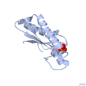
| - | == | + | ==Mad structure of the periplasmique domain of the Escherichia coli PAL protein== |
| - | + | <StructureSection load='1oap' size='340' side='right'caption='[[1oap]], [[Resolution|resolution]] 1.93Å' scene=''> | |
| - | [[ | + | == Structural highlights == |
| - | [[ | + | <table><tr><td colspan='2'>[[1oap]] is a 1 chain structure with sequence from [https://en.wikipedia.org/wiki/Escherichia_coli Escherichia coli]. Full crystallographic information is available from [http://oca.weizmann.ac.il/oca-bin/ocashort?id=1OAP OCA]. For a <b>guided tour on the structure components</b> use [https://proteopedia.org/fgij/fg.htm?mol=1OAP FirstGlance]. <br> |
| - | [[ | + | </td></tr><tr id='method'><td class="sblockLbl"><b>[[Empirical_models|Method:]]</b></td><td class="sblockDat" id="methodDat">X-ray diffraction, [[Resolution|Resolution]] 1.93Å</td></tr> |
| - | [ | + | <tr id='ligand'><td class="sblockLbl"><b>[[Ligand|Ligands:]]</b></td><td class="sblockDat" id="ligandDat"><scene name='pdbligand=SO4:SULFATE+ION'>SO4</scene></td></tr> |
| - | [ | + | <tr id='resources'><td class="sblockLbl"><b>Resources:</b></td><td class="sblockDat"><span class='plainlinks'>[https://proteopedia.org/fgij/fg.htm?mol=1oap FirstGlance], [http://oca.weizmann.ac.il/oca-bin/ocaids?id=1oap OCA], [https://pdbe.org/1oap PDBe], [https://www.rcsb.org/pdb/explore.do?structureId=1oap RCSB], [https://www.ebi.ac.uk/pdbsum/1oap PDBsum], [https://prosat.h-its.org/prosat/prosatexe?pdbcode=1oap ProSAT]</span></td></tr> |
| - | + | </table> | |
| - | + | == Function == | |
| - | + | [https://www.uniprot.org/uniprot/PAL_ECOLI PAL_ECOLI] Thought to play a role in bacterial envelope integrity. Very strongly associated with the peptidoglycan. | |
| - | [ | + | == Evolutionary Conservation == |
| - | [[ | + | [[Image:Consurf_key_small.gif|200px|right]] |
| - | [ | + | Check<jmol> |
| - | [[ | + | <jmolCheckbox> |
| - | [ | + | <scriptWhenChecked>; select protein; define ~consurf_to_do selected; consurf_initial_scene = true; script "/wiki/ConSurf/oa/1oap_consurf.spt"</scriptWhenChecked> |
| - | + | <scriptWhenUnchecked>script /wiki/extensions/Proteopedia/spt/initialview01.spt</scriptWhenUnchecked> | |
| + | <text>to colour the structure by Evolutionary Conservation</text> | ||
| + | </jmolCheckbox> | ||
| + | </jmol>, as determined by [http://consurfdb.tau.ac.il/ ConSurfDB]. You may read the [[Conservation%2C_Evolutionary|explanation]] of the method and the full data available from [http://bental.tau.ac.il/new_ConSurfDB/main_output.php?pdb_ID=1oap ConSurf]. | ||
| + | <div style="clear:both"></div> | ||
| + | <div style="background-color:#fffaf0;"> | ||
| + | == Publication Abstract from PubMed == | ||
| + | The peptidoglycan-associated lipoprotein (Pal) from Escherichia coli is part of the Tol--Pal multiprotein complex used by group A colicins to penetrate and kill cells. Pal homologues are found in many Gram-negative bacteria and the Tol--Pal system is thought to play a role in bacterial envelope integrity. The Pal protein comprises 152 amino acids. Crystals of the C-terminal 109-amino-acid fragment of the Pal protein have been produced. The crystals belong to the tetragonal space group I4(1), with unit-cell parameters a = b = 89.3, c = 67.2 A. There are two molecules in the asymmetric unit. Frozen crystals diffract to at least 2.8 A resolution using synchrotron radiation. Selenomethionine-substituted truncated Pal protein is currently being produced in order to use multiwavelength anomalous dispersion (MAD) for phasing. | ||
| - | + | Crystallization and preliminary crystallographic study of the peptidoglycan-associated lipoprotein from Escherichia coli.,Abergel C, Walburger A, Chenivesse S, Lazdunski C Acta Crystallogr D Biol Crystallogr. 2001 Feb;57(Pt 2):317-9. PMID:11173492<ref>PMID:11173492</ref> | |
| + | |||
| + | From MEDLINE®/PubMed®, a database of the U.S. National Library of Medicine.<br> | ||
| + | </div> | ||
| + | <div class="pdbe-citations 1oap" style="background-color:#fffaf0;"></div> | ||
| + | |||
| + | ==See Also== | ||
| + | *[[Pal|Pal]] | ||
| + | == References == | ||
| + | <references/> | ||
| + | __TOC__ | ||
| + | </StructureSection> | ||
| + | [[Category: Escherichia coli]] | ||
| + | [[Category: Large Structures]] | ||
| + | [[Category: Abergel C]] | ||
| + | [[Category: Bouveret E]] | ||
| + | [[Category: Claverie JM]] | ||
| + | [[Category: Walburger A]] | ||
Current revision
Mad structure of the periplasmique domain of the Escherichia coli PAL protein
| |||||||||||