This old version of Proteopedia is provided for student assignments while the new version is undergoing repairs. Content and edits done in this old version of Proteopedia after March 1, 2026 will eventually be lost when it is retired in about June of 2026.
Apply for new accounts at the new Proteopedia. Your logins will work in both the old and new versions.
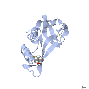
7rsa
From Proteopedia
(Difference between revisions)
| (8 intermediate revisions not shown.) | |||
| Line 1: | Line 1: | ||
| - | {{Seed}} | ||
| - | [[Image:7rsa.png|left|200px]] | ||
| - | + | ==STRUCTURE OF PHOSPHATE-FREE RIBONUCLEASE A REFINED AT 1.26 ANGSTROMS== | |
| - | + | <StructureSection load='7rsa' size='340' side='right'caption='[[7rsa]], [[Resolution|resolution]] 1.26Å' scene=''> | |
| - | You may | + | == Structural highlights == |
| - | or the | + | <table><tr><td colspan='2'>[[7rsa]] is a 1 chain structure with sequence from [https://en.wikipedia.org/wiki/Bos_taurus Bos taurus]. Full crystallographic information is available from [http://oca.weizmann.ac.il/oca-bin/ocashort?id=7RSA OCA]. For a <b>guided tour on the structure components</b> use [https://proteopedia.org/fgij/fg.htm?mol=7RSA FirstGlance]. <br> |
| - | + | </td></tr><tr id='method'><td class="sblockLbl"><b>[[Empirical_models|Method:]]</b></td><td class="sblockDat" id="methodDat">X-ray diffraction, [[Resolution|Resolution]] 1.26Å</td></tr> | |
| - | - | + | <tr id='ligand'><td class="sblockLbl"><b>[[Ligand|Ligands:]]</b></td><td class="sblockDat" id="ligandDat"><scene name='pdbligand=TBU:TERTIARY-BUTYL+ALCOHOL'>TBU</scene></td></tr> |
| - | + | <tr id='resources'><td class="sblockLbl"><b>Resources:</b></td><td class="sblockDat"><span class='plainlinks'>[https://proteopedia.org/fgij/fg.htm?mol=7rsa FirstGlance], [http://oca.weizmann.ac.il/oca-bin/ocaids?id=7rsa OCA], [https://pdbe.org/7rsa PDBe], [https://www.rcsb.org/pdb/explore.do?structureId=7rsa RCSB], [https://www.ebi.ac.uk/pdbsum/7rsa PDBsum], [https://prosat.h-its.org/prosat/prosatexe?pdbcode=7rsa ProSAT]</span></td></tr> | |
| + | </table> | ||
| + | == Function == | ||
| + | [https://www.uniprot.org/uniprot/RNAS1_BOVIN RNAS1_BOVIN] Endonuclease that catalyzes the cleavage of RNA on the 3' side of pyrimidine nucleotides. Acts on single stranded and double stranded RNA.<ref>PMID:7479688</ref> | ||
| + | == Evolutionary Conservation == | ||
| + | [[Image:Consurf_key_small.gif|200px|right]] | ||
| + | Check<jmol> | ||
| + | <jmolCheckbox> | ||
| + | <scriptWhenChecked>; select protein; define ~consurf_to_do selected; consurf_initial_scene = true; script "/wiki/ConSurf/rs/7rsa_consurf.spt"</scriptWhenChecked> | ||
| + | <scriptWhenUnchecked>script /wiki/extensions/Proteopedia/spt/initialview01.spt</scriptWhenUnchecked> | ||
| + | <text>to colour the structure by Evolutionary Conservation</text> | ||
| + | </jmolCheckbox> | ||
| + | </jmol>, as determined by [http://consurfdb.tau.ac.il/ ConSurfDB]. You may read the [[Conservation%2C_Evolutionary|explanation]] of the method and the full data available from [http://bental.tau.ac.il/new_ConSurfDB/main_output.php?pdb_ID=7rsa ConSurf]. | ||
| + | <div style="clear:both"></div> | ||
| + | <div style="background-color:#fffaf0;"> | ||
| + | == Publication Abstract from PubMed == | ||
| + | The structure of phosphate-free bovine ribonuclease A has been refined at 1.26-A resolution by a restrained least-squares procedure to a final R factor of 0.15. X-ray diffraction data were collected with an electronic position-sensitive detector. The final model consists of all atoms in the polypeptide chain including hydrogens, 188 water sites with full or partial occupancy, and a single molecule of 2-methyl-2-propanol. Thirteen side chains were modeled with two alternate conformations. Major changes to the active site include the addition of two waters in the phosphate-binding pocket, disordering of Gln-11, and tilting of the imidazole ring of His-119. The structure of the protein and of the associated solvent was extensively compared with three other high-resolution, refined structures of this enzyme. | ||
| - | + | Structure of phosphate-free ribonuclease A refined at 1.26 A.,Wlodawer A, Svensson LA, Sjolin L, Gilliland GL Biochemistry. 1988 Apr 19;27(8):2705-17. PMID:3401445<ref>PMID:3401445</ref> | |
| + | From MEDLINE®/PubMed®, a database of the U.S. National Library of Medicine.<br> | ||
| + | </div> | ||
| + | <div class="pdbe-citations 7rsa" style="background-color:#fffaf0;"></div> | ||
| - | + | ==See Also== | |
| - | + | *[[Ribonuclease 3D structures|Ribonuclease 3D structures]] | |
| - | + | == References == | |
| - | + | <references/> | |
| - | + | __TOC__ | |
| - | + | </StructureSection> | |
| - | == | + | |
| - | + | ||
| - | + | ||
| - | == | + | |
| - | + | ||
[[Category: Bos taurus]] | [[Category: Bos taurus]] | ||
| - | [[Category: | + | [[Category: Large Structures]] |
| - | + | [[Category: Gilliland GL]] | |
| - | [[Category: Gilliland | + | [[Category: Wlodawer A]] |
| - | [[Category: Wlodawer | + | |
| - | + | ||
| - | + | ||
Current revision
STRUCTURE OF PHOSPHATE-FREE RIBONUCLEASE A REFINED AT 1.26 ANGSTROMS
| |||||||||||