Interleukin receptor
From Proteopedia
(Difference between revisions)
| (4 intermediate revisions not shown.) | |||
| Line 1: | Line 1: | ||
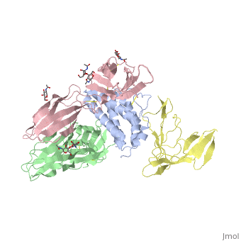
<StructureSection load='2b5i' size='340' side='right' caption='Glycosylated interleukin-2 receptor α (yellow), β (green), γ (pink) chains complex with interleukin-2 (grey), [[2b5i]]' scene=''> | <StructureSection load='2b5i' size='340' side='right' caption='Glycosylated interleukin-2 receptor α (yellow), β (green), γ (pink) chains complex with interleukin-2 (grey), [[2b5i]]' scene=''> | ||
| - | '''Interleukin receptors''' (ILR) are cytokine | + | __TOC__ |
| + | ==Function== | ||
| + | '''Interleukin receptors''' (ILR) are cytokine receptors belonging to the ILR of immunoglobulin family. which are classified into type I, type II and others. | ||
| + | *'''ILR type I''' are cell surface receptors which recognize cytokines with four α-helical strand. binding interleukin-1 (IL1) transmitting its inflammatory effect. | ||
| + | *'''ILR type II''' are similar to type I but lack the former’s signature sequence: WXSWS. They suppresses IL1 activity. Another suppressor of IL1R activity is the interleukin-1 receptor antagonist (IL1R1A). | ||
| - | '''TYPE-1 INTERLEUKIN-1 RECEPTOR COMPLEXED WITH INTERLEUKIN-1 BETA''' | + | See also: [[Cytokine receptors]]. |
| + | |||
| + | =='''TYPE-1 INTERLEUKIN-1 RECEPTOR COMPLEXED WITH INTERLEUKIN-1 BETA''' == | ||
[http://en.wikipedia.org/wiki/Interleukin-1_receptor Interleukin-1 receptor] complex with ligand and go through the plasma membrane. <scene name='57/571319/Scene_2/2'> Type 1 Interleukin-1 receptor complex with Interleukin-1 beta</scene> 3D structure is showing here. Ribbon diagram of s-IL 1R complex to IL-1β. The complex has approximate dimensions of 97Å × 52Å ×35Å with one s-IL1R molecule wrapping around the IL-1β molecule with 1:1 ratio. In the <scene name='57/571319/Scene_2/3'>complex</scene>, domain 3 provides a 'lid' which covers most of the top of the IL-1β β-barrel, whereas domains 1 and 2 from a groove which binds to the lower rim of the barrel. Here, Domains 1,2 and 3 of s-IL 1R are colored light, medium and dark blue, respectively. IL-1β is yellow, with site A residues in green and site B residues in red. The structure is oriented so that the carboxy terminus of s-IL 1R and the cell membrane are at the bottom of the picture. | [http://en.wikipedia.org/wiki/Interleukin-1_receptor Interleukin-1 receptor] complex with ligand and go through the plasma membrane. <scene name='57/571319/Scene_2/2'> Type 1 Interleukin-1 receptor complex with Interleukin-1 beta</scene> 3D structure is showing here. Ribbon diagram of s-IL 1R complex to IL-1β. The complex has approximate dimensions of 97Å × 52Å ×35Å with one s-IL1R molecule wrapping around the IL-1β molecule with 1:1 ratio. In the <scene name='57/571319/Scene_2/3'>complex</scene>, domain 3 provides a 'lid' which covers most of the top of the IL-1β β-barrel, whereas domains 1 and 2 from a groove which binds to the lower rim of the barrel. Here, Domains 1,2 and 3 of s-IL 1R are colored light, medium and dark blue, respectively. IL-1β is yellow, with site A residues in green and site B residues in red. The structure is oriented so that the carboxy terminus of s-IL 1R and the cell membrane are at the bottom of the picture. | ||
| - | '''STRUCTURE OF THE INTERLEUKIN-1BETA SIGNALING COMPLEX''' | + | =='''STRUCTURE OF THE INTERLEUKIN-1BETA SIGNALING COMPLEX'''== |
[[Image:Ternary complex paradigm.png||250px|right|]] | [[Image:Ternary complex paradigm.png||250px|right|]] | ||
Current revision
| |||||||||||