ATPase
From Proteopedia
(Difference between revisions)
| (3 intermediate revisions not shown.) | |||
| Line 15: | Line 15: | ||
ATPase domains include metal-binding domain (MBD) and nucleotide-binding domain (NBD). For more details see:<br /> | ATPase domains include metal-binding domain (MBD) and nucleotide-binding domain (NBD). For more details see:<br /> | ||
* '''ATPase RavA''' participates in the pathway which response to ahminoglycosides under anaerobic conditions and cell membrane regulation<ref>PMID:36127320</ref>. <br /> | * '''ATPase RavA''' participates in the pathway which response to ahminoglycosides under anaerobic conditions and cell membrane regulation<ref>PMID:36127320</ref>. <br /> | ||
| - | *Cu transporting ATPase are in [[P(1B)-Type Cu(I) Transporting ATPases ATP7A and ATP7B]].<br /> | + | * '''ATPase InvC''' energizes the apparatus needed for the entry of ''Salmonella typhimurium'' into mammalian cells<ref>PMID:8045880</ref>. <br /> |
| - | *Na/K transporting ATPase are in [[Sodium-Potassium ATPase]].<br /> | + | * '''Peroxisomal ATPase''' Pex1/Pex6 is essential for peroxisome formation<ref>PMID:37741838</ref>. <br /> |
| - | *H/K transporting ATPase are in [[Esomeprazole and H+/K+ - ATPase Interaction]].<br /> | + | * '''INO8o ATPase''' is a component of the chromatin remodelling complex<ref>PMID:19062292</ref>. <br /> |
| - | *Transitional endoplasmic reticulum ATPase are in [[Valosin Containing Protein D120]].<br /> | + | *'''Cu transporting ATPase''' are in [[P(1B)-Type Cu(I) Transporting ATPases ATP7A and ATP7B]].<br /> |
| - | *Central stalk in F(1)-ATPase is described in [[A-ATP Synthase]]<br /> | + | *'''Na/K transporting ATPase''' are in [[Sodium-Potassium ATPase]].<br /> |
| + | *'''H/K transporting ATPase''' are in [[Esomeprazole and H+/K+ - ATPase Interaction]].<br /> | ||
| + | *'''Transitional endoplasmic reticulum ATPase''' are in [[Valosin Containing Protein D120]].<br /> | ||
| + | *'''Central stalk in F(1)-ATPase''' is described in [[A-ATP Synthase]]<br /> | ||
*[[Journal:JSB:1|RuvBL1/RuvBL2 complex (ATPase)]]<br /> | *[[Journal:JSB:1|RuvBL1/RuvBL2 complex (ATPase)]]<br /> | ||
| - | *For Ter-ATPase see [[Valosin Containing Protein D120]] | ||
Current revision
| |||||||||||
Contents |
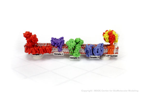
3D Printed Physical Model of ATP Synthase
Shown below is a 3D printed physical model of the Respiration Electron Transport Chain. Complex I is colored red, complex II is purple, complex III is green, complex IV is blue and the atp synthase protein is colored orange, yellow and red.

The MSOE Center for BioMolecular Modeling
The MSOE Center for BioMolecular Modeling uses 3D printing technology to create physical models of protein and molecular structures, making the invisible molecular world more tangible and comprehensible. To view more protein structure models, visit our Model Gallery.
3D Structures of ATPase
References
- ↑ Rappas M, Niwa H, Zhang X. Mechanisms of ATPases--a multi-disciplinary approach. Curr Protein Pept Sci. 2004 Apr;5(2):89-105. doi: 10.2174/1389203043486874. PMID:15078220 doi:http://dx.doi.org/10.2174/1389203043486874
- ↑ Neupane P, Bhuju S, Thapa N, Bhattarai HK. ATP Synthase: Structure, Function and Inhibition. Biomol Concepts. 2019 Mar 7;10(1):1-10. doi: 10.1515/bmc-2019-0001. PMID:30888962 doi:http://dx.doi.org/10.1515/bmc-2019-0001
- ↑ Abrahams JP, Leslie AG, Lutter R, Walker JE. Structure at 2.8 A resolution of F1-ATPase from bovine heart mitochondria. Nature. 1994 Aug 25;370(6491):621-8. PMID:8065448 doi:http://dx.doi.org/10.1038/370621a0
- ↑ Dupont C, Viljoen A, Thomas S, Roquet-Banères F, Herrmann JL, Pethe K, Kremer L. Bedaquiline Inhibits the ATP Synthase in Mycobacterium abscessus and Is Effective in Infected Zebrafish. Antimicrob Agents Chemother. 2017 Oct 24;61(11):e01225-17. PMID:28807917 doi:10.1128/AAC.01225-17
- ↑ Chan H, Babayan V, Blyumin E, Gandhi C, Hak K, Harake D, Kumar K, Lee P, Li TT, Liu HY, Lo TC, Meyer CJ, Stanford S, Zamora KS, Saier MH Jr. The p-type ATPase superfamily. J Mol Microbiol Biotechnol. 2010;19(1-2):5-104. doi: 10.1159/000319588. Epub 2010, Oct 20. PMID:20962537 doi:http://dx.doi.org/10.1159/000319588
- ↑ Plesner L. Ecto-ATPases: identities and functions. Int Rev Cytol. 1995;158:141-214. doi: 10.1016/s0074-7696(08)62487-0. PMID:7721538 doi:http://dx.doi.org/10.1016/s0074-7696(08)62487-0
- ↑ Felix J, Bumba L, Liesche C, Fraudeau A, Rébeillé F, El Khoury JY, Huard K, Gallet B, Moriscot C, Kleman JP, Duhoo Y, Jessop M, Kandiah E, Barras F, Jouhet J, Gutsche I. The AAA+ ATPase RavA and its binding partner ViaA modulate E. coli aminoglycoside sensitivity through interaction with the inner membrane. Nat Commun. 2022 Sep 20;13(1):5502. PMID:36127320 doi:10.1038/s41467-022-32992-9
- ↑ Eichelberg K, Ginocchio CC, Galán JE. Molecular and functional characterization of the Salmonella typhimurium invasion genes invB and invC: homology of InvC to the F0F1 ATPase family of proteins. J Bacteriol. 1994 Aug;176(15):4501-10. PMID:8045880 doi:10.1128/jb.176.15.4501-4510.1994
- ↑ Rüttermann M, Koci M, Lill P, Geladas ED, Kaschani F, Klink BU, Erdmann R, Gatsogiannis C. Structure of the peroxisomal Pex1/Pex6 ATPase complex bound to a substrate. Nat Commun. 2023 Sep 23;14(1):5942. PMID:37741838 doi:10.1038/s41467-023-41640-9
- ↑ Conaway RC, Conaway JW. The INO80 chromatin remodeling complex in transcription, replication and repair. Trends Biochem Sci. 2009 Feb;34(2):71-7. PMID:19062292 doi:10.1016/j.tibs.2008.10.010
Proteopedia Page Contributors and Editors (what is this?)
Michal Harel, Wayne Decatur, Alexander Berchansky, Mark Hoelzer, Karsten Theis, Jaime Prilusky

