From Proteopedia
(Difference between revisions)
proteopedia linkproteopedia link
|
|
| Line 1: |
Line 1: |
| - | <StructureSection load='7jzl' size='350' frame='true' side='right' caption='SARS-CoV-2 Spike Protein Bound to Minibinders (PDB 7jzl)' scene='10/1075220/Monomerwithminibinder/4'> | + | <StructureSection load='7jzl' size='350' frame='true' side='right' caption='SARS-CoV-2 Spike Protein (7JZL): A trimer responsible for interacting with host ACE2 receptors to deliver the virus into host cells.' scene='10/1078099/Spike_protein/1'> |
| | | | |
| | ==Introduction== | | ==Introduction== |
Revision as of 01:09, 14 April 2025
|
Introduction
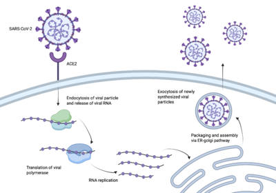 Figure 1: The process of the SARS-CoV-2 virus entering human cells. The image was created using the program biorender. SARS-CoV-2 better known as Covid-19 sent the world into a global pandemic due to its rapid transmission and infection [1]. In order to create a vaccine, it was essential to understand how SARS-CoV-2 infected us.
The enzyme angiotensin converting enzyme 2 is attached to our cell membranes and then can be bound to a receptor binding domain [2]. The virus, SARS-CoV-2, enters our bodies containing a spike protein protruding from the viral membrane. The RBD on the end of the spike protein giving SARS-CoV-2 the ability to enter our host cells [3]. Once the spike protein has access to our host cells, it is able to further infect our cells and spread the virus throughout our bodies, causing us to get sick. Figure 1 demonstrates the path SARS-CoV-2 takes to get into our cells. In order to create an effective vaccine, the pathway between the spike protein and the RBD needed to be interrupted.
Vaccines
A vaccine activates the body’s immune system by containing weakened parts of viruses. This activation stimulates the production of antibodies. then compete with the ACE2 protein for the binding of viral spike proteins. In the case of SARS-CoV-2 spike proteins, once they enter the body they <scenename='10/1077473/Antibody/4'>bind</scene>
to the Fab fragment of the antibody, a 90% neutralizing response for targeting the RBD is created. In this scene only the Fab fragment is being shown due to the fact that the Fab fragment is the part of the antibody that interacts with the RBD. With this being said, this method of treatment is difficult for long term use due to the evolution of the viral cells [4].
Protein Inhibitor Development
Protein inhibitors were thought of as a new idea for creating vaccines due to their smaller size and better stability compared to antibody vaccines[5]. These protein inhibitors, also referred to as mini-binders, interact with the receptor binding domain of the spike protein,
The Process of Discovery
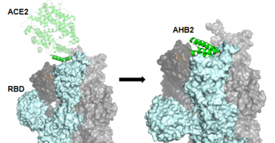 Figure 2: The use of the Rosetta Blueprint protein design to create the AHB2 inhibitor. The first mini-binder to be created to combat COVID-19 is called AHB2. In order to ensure that the mini-binder would bind to the same RBD that the ACE2 was bound to, AHB2 was designed by looking at the specific sequence of ACE2 to find the alpha-helix that makes interactions with the spike receptor binding domain. This design process is referred to as the Rosetta Blueprint protein design. Figure 2 shows the RBD trimer with one part of ACE2 being used for the reference alpha helix to create AHB2. [5].
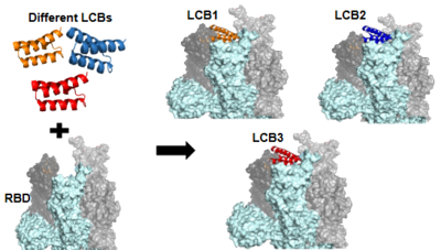 Figure 3:The use of the De Novo protein design to create the LCB1 and LCB3 inhibitors. As the AHB2 inhibitors were tested and found to be effective, it was then time to manipulate the mini-binders to create a more effective vaccine. A rotamer interaction field docking with in silico mini-proteins were used by using a scaffold library to generate binders to more distinct regions of the RBD surface [5]. This method is known as the de novo protein design and it is how the LCB1 and LCB3 mini-binders were created. Figure 3 shows the difference LCBs pulled from the scaffold library to create the different LCB inhibitors.
Stability
One of the most important findings with these DeNovo proteins is their high stability, allowing for less delicate forms of administration.
,
, and
|
Binding Site and Interactions
In order to best target the RBD of the spike protein, the minibinders reveal a wide range of interactions to compete with .
The first design method, Rosetta, created AHB2 based on the single interacting helix of ACE2. With , we see two alpha helices mimicking ACE2. Hydrogen bonding interactions between N36, D11, K43, E41, and E30 of the minibinder interact with residues K417, R403, Y449, Q493, and N487, respectively, in the spike protein.
De novo designed proteins, as discussed previously, focused on computational design to determine residues best able to interact with the spike protein. We will focus on LCB1 and LCB3. reveals hydrogen bonding between D30 of the minibinder and both K417 and R403 of the spike protein, in addition to D17 and R14 of the minibinder interacting with Q493 of the spike protein. Similarly, reveals hydrogen bonding between D11 of the minibinder to K417 and R403 of the spike protein.
Within all four binding sites, we see two conserved residues throughout: K417 and Q493. This finding reveals the importance of these residues in both binding and stability.
Furthermore, an interesting difference between the two design methods is the location of hydrogen bonding interactions. In AHB2, similar to ACE2, we see interactions spanning the entirety of the helices in contact with the spike protein. De novo design, on the other hand, reveals the main hydrogen bonding interactions occurring within the center residues of the helices.
Figure 4 shows the sequence comparison between the interacting helix of ACE2 and the helices of the three minibinders.
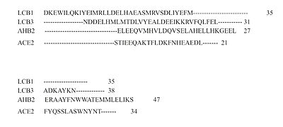
Figure 4: The sequence differences between ACE2, AHB2, LCB1 and LCB3.
Protein Inhibitor Effectiveness
Limitations
References
Cao, L., Goreshnik, I., Coventry, B., Case, J.B., Miller, L., Kozodoy, L., Chen, R.E., Carter, L., Walls, A.C., Park, Y., Strauch, E., Stewart, L., Diamond, M.S., Veesler, D., & Baker, D. De novo design of picomolar SARS-CoV-2 mini protein inhibitors. Science 370, 426-431 (2020). https://doi.org/10.1126/science.abd9909
https://www.who.int/europe/emergencies/situations/covid-19
https://pmc.ncbi.nlm.nih.gov/articles/PMC9786537/#:~:text=The%20receptor%2Dbinding%20domain%20(RBD,that%20initiates%20the%20viral%20transmission.
https://www.nature.com/articles/s41580-021-00418-x#citeas
https://www.science.org/doi/10.1126/science.abd9909
Zhang, Haoran et al. Advances in developing ACE2 derivatives against SARS-CoV-2. The Lancet Microbe, Volume 4, Issue 5, e369 - e378 (2023). https://doi.org/10.1016/S2666-5247(23)00011-3
https://www.cdc.gov/vaccines/basics/explaining-how-vaccines-work.html\
https://en.wikipedia.org/wiki/Vaccine
PDB Files
[1]https://www.rcsb.org/structure/7UHB
Student Contributors
- Giavanna Yowell
- Shea Bailey
- Matthew Pereira


