We apologize for Proteopedia being slow to respond. For the past two years, a new implementation of Proteopedia has been being built. Soon, it will replace this 18-year old system. All existing content will be moved to the new system at a date that will be announced here.
Sandbox 4
From Proteopedia
(Difference between revisions)
| Line 1: | Line 1: | ||
| - | {{Seed}} | ||
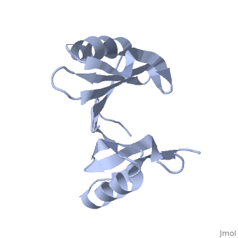
| - | [[Image:1tsj.png|left|200px]] | ||
| - | |||
| - | <!-- | ||
| - | The line below this paragraph, containing "STRUCTURE_1tsj", creates the "Structure Box" on the page. | ||
| - | You may change the PDB parameter (which sets the PDB file loaded into the applet) | ||
| - | or the SCENE parameter (which sets the initial scene displayed when the page is loaded), | ||
| - | or leave the SCENE parameter empty for the default display. | ||
| - | --> | ||
| - | |||
| - | =Crystal structure of protein from Staphylococcus aureus= | ||
| - | |||
| - | PDB entry 1tsj refers to a hypothetical protein of 139 residues which is a predicted dimer<ref> Henrick, K., and Thornton, J.M. (1998). PQS: a protein quaternary structure file server. Trends Biochem. Sci. 23, 358- 361.</ref> and predicted as a cytoplasmic protein.<ref> Bhasin, M., Garg, A. and Raghava, GPS (2005) PSLpred: prediction of subcellular localization of bacterial proteins. Bioinformatics. 21, 2522-2524. | ||
| - | Gardy JL, Spencer C, Wang K, Ester M, Tusn?dy GE, Simon I, Hua S, deFays K, Lambert C, Nakai K, Brinkman FS. (2003) PSORT-B: improving protein subcellular localization prediction for Gram-negative bacteria. Nucleic Acids Res. 31, 3613-3617. | ||
| - | Z. Lu, D. Szafron, R. Greiner, P. Lu, D.S. Wishart, B. Poulin, J. Anvik, C. Macdonell, and R. Eisner. (2003). Predicting Subcellular Localization of Proteins using Machine-Learned Classifiers. Bioinformatics. 20, 547-556. | ||
| - | Hua S, Sun Z. (2001). Support vector machine approach for protein subcellular localization prediction. Bioinformatics. 17, 721-728.</ref> | ||
| - | |||
The protein is associated with Pfam<ref>Finn R, Griffiths-Jones S, Bateman A. (2003). Identifying protein domains with the Pfam database. Curr Protoc Bioinformatics. Chapter 2: Unit 2.5.</ref> entry PF06983 of 3-demethylubiquinone-9 3-methyltransferases. | The protein is associated with Pfam<ref>Finn R, Griffiths-Jones S, Bateman A. (2003). Identifying protein domains with the Pfam database. Curr Protoc Bioinformatics. Chapter 2: Unit 2.5.</ref> entry PF06983 of 3-demethylubiquinone-9 3-methyltransferases. | ||
| - | + | <applet load='1tsj' size='400' frame='true' align='right' caption='1tsj, resolution 2.60Å' /> <scene name='1tsj/Evolutionary_consevation_1tsj/1'>highly preserved</scene> and negatively charged. | |
| - | + | ||
| - | + | ||
| - | + | ||
| - | + | ||
| - | + | ||
| - | + | ||
| - | + | ||
| - | + | ||
| - | + | ||
| - | + | ||
| - | + | ||
| - | + | ||
| - | + | ||
| - | + | ||
| - | + | ||
| - | + | ||
| - | + | ||
<center>{{Template:ColorKey_ConSurf}}</center> | <center>{{Template:ColorKey_ConSurf}}</center> | ||
| Line 46: | Line 12: | ||
==References== | ==References== | ||
<references/> | <references/> | ||
| - | |||
| - | [[Category: Staphylococcus aureus subsp. aureus]] | ||
| - | [[Category: Burley, S K.]] | ||
| - | [[Category: Gorman, J.]] | ||
| - | [[Category: Min, T.]] | ||
| - | [[Category: NYSGXRC, New York Structural GenomiX Research Consortium.]] | ||
| - | [[Category: Shapiro, L.]] | ||
| - | [[Category: Conserved hypothetical protein]] | ||
| - | [[Category: Crystal structure]] | ||
| - | [[Category: New york structural genomics consortium]] | ||
| - | [[Category: New york structural genomix research consortium]] | ||
| - | [[Category: Nysgxrc]] | ||
| - | [[Category: Protein structure initiative]] | ||
| - | [[Category: Psi]] | ||
| - | [[Category: Structural genomic]] | ||
Revision as of 16:54, 18 February 2009
The protein is associated with Pfam[1] entry PF06983 of 3-demethylubiquinone-9 3-methyltransferases.
|

The potential substrate S-adenosyl-L-methionine has positive formal charge which fits the negative electrostatic potential in the potential catalytic area. The alternative substrate found by the structural homology methylglyoxal is not charged and thus less probable to attach to the potential catalytic area.
It seems more likely that Q8NX24 belongs to the Glyoxalase/bleomycin resistance protein/dioxygenase superfamily and functions as a demethylubiquinone-9 3-methyltransferase protein.
References
- ↑ Finn R, Griffiths-Jones S, Bateman A. (2003). Identifying protein domains with the Pfam database. Curr Protoc Bioinformatics. Chapter 2: Unit 2.5.
