We apologize for Proteopedia being slow to respond. For the past two years, a new implementation of Proteopedia has been being built. Soon, it will replace this 18-year old system. All existing content will be moved to the new system at a date that will be announced here.
Sandbox 107
From Proteopedia
(Difference between revisions)
| (7 intermediate revisions not shown.) | |||
| Line 1: | Line 1: | ||
<table style="background-color:#ffffc0" cellpadding="8" width="95%" border="0"><tr><td>Please do NOT make changes to this Sandbox until after December 15, 2009. Sandboxes 85-115 are reserved until then for use by the Biochemistry 451 class at Capital University taught by Prof. Jens Hemmingsen (jhemming@capital.edu).</td></tr> | <table style="background-color:#ffffc0" cellpadding="8" width="95%" border="0"><tr><td>Please do NOT make changes to this Sandbox until after December 15, 2009. Sandboxes 85-115 are reserved until then for use by the Biochemistry 451 class at Capital University taught by Prof. Jens Hemmingsen (jhemming@capital.edu).</td></tr> | ||
| - | + | {{Seed}} | |
| + | [[Image:3e3f.jpg|left|200px]] | ||
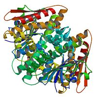
| - | {{STRUCTURE_1a6m | PDB= | + | This is "LueT", it is a transmembrane protein. |
| - | <scene name=' | + | |
| - | <scene name=' | + | {{STRUCTURE_1a6m | PDB=3f3e | SCENE= }} |
| - | <scene name=' | + | |
| - | <scene name=' | + | <scene name='Sandbox_100/Original_view/1'>Original view</scene> |
| + | |||
| + | <scene name='Sandbox_100/Leut_bound_with_tryptophan/1'>Hydrophobic/polar groups</scene> | ||
| + | |||
| + | <scene name='Sandbox_100/Leut_with_trp_bound/4'>Inhibitor bound</scene> | ||
| + | |||
| + | <scene name='Sandbox_100/Leut/1'>Rocket view of LeuT</scene> | ||
Current revision
| Please do NOT make changes to this Sandbox until after December 15, 2009. Sandboxes 85-115 are reserved until then for use by the Biochemistry 451 class at Capital University taught by Prof. Jens Hemmingsen (jhemming@capital.edu). | |||||||||
| |||||||||
| 1a6m, resolution 1.00Å () | |||||||||
|---|---|---|---|---|---|---|---|---|---|
| Ligands: | , , | ||||||||
| |||||||||
| |||||||||
| |||||||||
| Resources: | FirstGlance, OCA, PDBsum, RCSB | ||||||||
| Coordinates: | save as pdb, mmCIF, xml | ||||||||


