|
|
| (3 intermediate revisions not shown.) |
| Line 1: |
Line 1: |
| - | == M2 Proton Channel from ''Influenza'' A Virus == | + | == Chloride Ion Channel == |
| - | <applet load='1nyj' size='300' frame='true' align='right' caption='The | + | |
| - | closed state structure of M2 protein H+ channel by solid state NMR
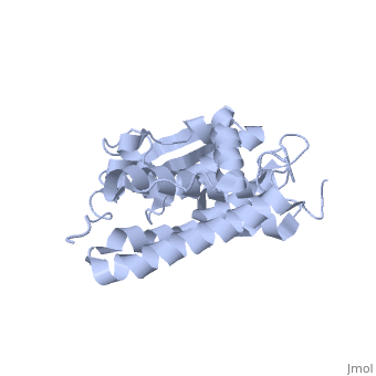
| + | Crystal structure of a soluble form of CLIC1. An intracellular chloride ion channel |
| - | spectroscopy. [Stouffer et al, 2008]' />
| + | |
| - | | + | <applet load='1k0o' size='300' frame='true' align='right' caption='Insert caption here' /> |
| - | == Background ==
| + | |
| - | The M2 proton channel is a key protein that leads to viral infection.<ref
| + | CLIC1 (NCC27) is a member of the highly conserved class of chloride ion channels that exists in both soluble and integral membrane forms. Purified CLIC1 can integrate into synthetic lipid bilayers forming a chloride channel with similar properties to those observed in vivo. The structure of the soluble form of CLIC1 has been determined at 1.4-A resolution. The protein is monomeric and structurally homologous to the glutathione S-transferase superfamily, and it has a redox-active site resembling glutaredoxin. The structure of the complex of CLIC1 with glutathione shows that glutathione occupies the redox-active site, which is adjacent to an open, elongated slot lined by basic residues. Integration of CLIC1 into the membrane is likely to require a major structural rearrangement, probably of the N-domain (residues 1-90), with the putative transmembrane helix arising from residues in the vicinity of the redox-active site. The structure indicates that CLIC1 is likely to be controlled by redox-dependent processes. |
| - | name="Takeuchi" /> The M2 proton channel acidifies the viron which allows
| + | Crystal structure of a soluble form of the intracellular chloride ion channel CLIC1 (NCC27) at 1.4-A resolution. |
| - | the viral matrix protein (M1) to disassociate from the ribonucleoprotein
| + | |
| - | (RNP).<ref name="Wu">PMID:12972147 </ref> This allows the RNP to be
| + | |
| - | transported to the nucleus of the cell.<ref name="Wu" /> Several recent
| + | == About this Structure == |
| - | studies have looked at the effects of <scene
| + | |
| - | name='User:Sarah_Henke/Sandbox_1/Amantadine/1'>amantadine</scene><ref
| + | |
| - | name="Stouffer">PMID:18235504 </ref> and <scene
| + | |
| - | name='User:Sarah_Henke/Sandbox_1/Rimantadine/1'>rimantadine</scene><ref
| + | 1K0O is a 2 chains structure of sequences from Homo sapiens. Full crystallographic information is available from OCA. |
| - | name="Schnell">PMID:18235503 </ref> on inhibiting the transfer of protons
| + | [edit] |
| - | through the M2 channel.<ref name="Stouffer" /> It has been found that M2 is
| + | |
| - | resistant to these two drugs in 90% of humans, birds and pigs.<ref
| + | |
| - | name="Stouffer" /> Understanding the structure and function of this proton
| + | |
| - | channel is necessary in solving the resistance problem.<ref name="Stouffer" | + | |
| - | /> The <scene name='User:Sarah_Henke/Sandbox_1/3bkd/2'>crystal
| + | |
| - | structure</scene> of the M2 proton channel influenza A virus was solved in | + | |
| - | 2008 (PBD: 3bkd).<ref name="Stouffer" />
| + | |
| - | | + | |
| - | == Structure ==
| + | |
| - | The M2 proton channel from influenza A is 97 amino acid residues and forms a | + | |
| - | 24-residue N-terminal extracellular domain, a 19-residue trans-membrane
| + | |
| - | domain, and a 54-residue C-terminal cytoplasmic domain.<ref name="Wu" /> The
| + | |
| - | 19-residue TM domain forms the highly selective proton channel.<ref
| + | |
| - | name="Takeuchi">PMID:12972149 </ref> Circular dichroism spectra has shown
| + | |
| - | the TM domain to form an <scene | + | |
| - | name='User:Sarah_Henke/Sandbox_1/Momomer/2'>α-helix</scene> that spans the
| + | |
| - | membrane.<ref name="Wu" /> By analytical ultracentrifugation, the TM domain
| + | |
| - | is found to form <scene | + | |
| - | name='User:Sarah_Henke/Sandbox_1/Alpha_hlix/1'>homotetramers</scene> which
| + | |
| - | contains four identical α-helices.<ref name="Takeuchi" /> Secondary
| + | |
| - | structure is color coded by {{Template:ColorKey_Helix}}. When viewed in the
| + | |
| - | <scene name='User:Sarah_Henke/Sandbox_1/N_to_c/1'>N->C color coding</scene>
| + | |
| - | the <FONT COLOR="blue">'''N-terminus'''</FONT> is located near the
| + | |
| - | extracellular side of the membrane while the <FONT
| + | |
| - | COLOR="red">'''C-terminus'''</FONT> is located near the cytosolic side of
| + | |
| - | the membrane. This tetrameric bundle of the TM domain is found by NMR to be
| + | |
| - | tilted by 25-38° from the channel axis.<ref name="Takeuchi" /> The trameric
| + | |
| - | helices form a left-handed bundle that resembles a truncated cone.<ref
| + | |
| - | name="Stouffer" /> The TM helicies are arranged around the channel pore with
| + | |
| - | an approximate four-fold rotational symmetry.<ref name="Takeuchi" />
| + | |
| - | | + | |
| - | == Central Cavity ==
| + | |
| - | <applet load='3bkd' size='300' frame='true' align='right' caption='High
| + | |
| - | resolution Crystal structure of Transmembrane domain of M2 protein.
| + | |
| - | [Stouffer et al, 2008]' />
| + | |
| - | The hydrophilic residues in each α-helix monomer are oriented towards the
| + | |
| - | pore lumen.<ref name="Wu" /> The <scene
| + | |
| - | name='User:Sarah_Henke/Sandbox_1/Hydrophobic/1'>hydrophobic</scene> residues
| + | |
| - | will be in contact with the membrane (Color code=
| + | |
| - | {{Template:ColorKey_Hydrophobic}}). Most of the residues in the M2 channel
| + | |
| - | are hydrophobic except Ser31 Gly34, and His37.<ref name="Wu" /> The central
| + | |
| - | cavity of the M2 channel is a water-filled pore that is interrupted at
| + | |
| - | residue <scene name='User:Sarah_Henke/Sandbox_1/His_37/1'>His37</scene> in
| + | |
| - | each monomer.<ref name="Lear">PMID:12972146 </ref> Mutagenesis studies have
| + | |
| - | found that the residues facing the pore are Val27, Ala30, Ser31, Gly34,
| + | |
| - | His37, Leu38, and Trp41.<ref name="Wu" /> Residues His37 and <scene
| + | |
| - | name='User:Sarah_Henke/Sandbox_1/Trp41/1'>Trp41</scene> play a key role in
| + | |
| - | the gating mechanism and the selectivity filter.<ref name="Wu" /> In the
| + | |
| - | <scene name='User:Sarah_Henke/Sandbox_1/Trphis/1'>closed state</scene>, the
| + | |
| - | His37 and Trp41 residues block the channel, preventing proton conductance.
| + | |
| - | | + | |
| - | == pH Gating ==
| + | |
| - | The M2 channel is low-pH gated and has a 50-fold increase in proton
| + | |
| - | conductance when the pH drops from 8.2 down to 4.2.<ref name="Wu" /> The His
| + | |
| - | side chain, a five-membered ring, has two nitrogen atoms.<ref
| + | |
| - | name="Takeuchi" /> At a pH of 7 or higher, only one nitrogen is protonated
| + | |
| - | in the neutral imidazole form.<ref name="Takeuchi" /> However, at a pH of
| + | |
| - | about 5.8, the second nitrogen becomes protonated and forms the cationic
| + | |
| - | imidazolium form.<ref name="Takeuchi" /> This protonation causes the His
| + | |
| - | residues in each monomer to move away from each other due to electrostatic
| + | |
| - | repulsion.<ref name="Takeuchi" /><ref name="Lear" /><ref name="Wu" /> This
| + | |
| - | allows the channel to open and allow water and protons to pass through.<ref
| + | |
| - | name="Lear" /><ref name="Wu" />
| + | |
| - | | + | |
| - | == Selectivity ==
| + | |
| - | The selectivity of this channel is based on the binding of a proton to the
| + | |
| - | His37 residues in each of the α-helices in the tetramer.<ref name="Takeuchi"
| + | |
| - | /><ref name="Lear" /><ref name="Wu" /> Without protons binding to the His
| + | |
| - | residues, the channel would not open. Also, because there is an excess
| + | |
| - | amount of protons when the pH is in acidic conditions on the extracellular
| + | |
| - | side of the membrane, a proton gradient is formed. Once the channel is open,
| + | |
| - | the protons move with their gradient to the cytosolic side of the membrane.
| + | |
| - | These protons are then used to acidify the viron.<ref name="Wu" />
| + | |
| - | | + | |
| - | | + | |
| | == References == | | == References == |
| - | <references /> | + | |
| | + | <references/> |
| | + | |
| | + | <ref>PMID:#11551966</ref> |
| | + | |
| | + | Harrop SJ, DeMaere MZ, Fairlie WD, Reztsova T, Valenzuela SM, Mazzanti M, Tonini R, Qiu MR, Jankova L, Warton K, Bauskin AR, Wu WM, Pankhurst S, Campbell TJ, Breit SN, Curmi PM. Crystal structure of a soluble form of the intracellular chloride ion channel CLIC1 (NCC27) at 1.4-A resolution. J Biol Chem. 2001 Nov 30;276(48):44993-5000. Epub 2001 Sep 10. PMID:11551966 |
Crystal structure of a soluble form of CLIC1. An intracellular chloride ion channel
CLIC1 (NCC27) is a member of the highly conserved class of chloride ion channels that exists in both soluble and integral membrane forms. Purified CLIC1 can integrate into synthetic lipid bilayers forming a chloride channel with similar properties to those observed in vivo. The structure of the soluble form of CLIC1 has been determined at 1.4-A resolution. The protein is monomeric and structurally homologous to the glutathione S-transferase superfamily, and it has a redox-active site resembling glutaredoxin. The structure of the complex of CLIC1 with glutathione shows that glutathione occupies the redox-active site, which is adjacent to an open, elongated slot lined by basic residues. Integration of CLIC1 into the membrane is likely to require a major structural rearrangement, probably of the N-domain (residues 1-90), with the putative transmembrane helix arising from residues in the vicinity of the redox-active site. The structure indicates that CLIC1 is likely to be controlled by redox-dependent processes.
Crystal structure of a soluble form of the intracellular chloride ion channel CLIC1 (NCC27) at 1.4-A resolution.
1K0O is a 2 chains structure of sequences from Homo sapiens. Full crystallographic information is available from OCA.
[edit]
Harrop SJ, DeMaere MZ, Fairlie WD, Reztsova T, Valenzuela SM, Mazzanti M, Tonini R, Qiu MR, Jankova L, Warton K, Bauskin AR, Wu WM, Pankhurst S, Campbell TJ, Breit SN, Curmi PM. Crystal structure of a soluble form of the intracellular chloride ion channel CLIC1 (NCC27) at 1.4-A resolution. J Biol Chem. 2001 Nov 30;276(48):44993-5000. Epub 2001 Sep 10. PMID:11551966