This old version of Proteopedia is provided for student assignments while the new version is undergoing repairs. Content and edits done in this old version of Proteopedia after March 1, 2026 will eventually be lost when it is retired in about June of 2026.
Apply for new accounts at the new Proteopedia. Your logins will work in both the old and new versions.
2wqd
From Proteopedia
(Difference between revisions)
| (10 intermediate revisions not shown.) | |||
| Line 1: | Line 1: | ||
| - | {{Seed}} | ||
| - | [[Image:2wqd.png|left|200px]] | ||
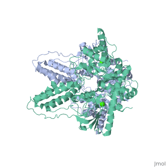
| - | < | + | ==Crystal structure of enzyme I of the phosphoenolpyruvate:sugar phosphotransferase system in the dephosphorylated state== |
| - | + | <StructureSection load='2wqd' size='340' side='right'caption='[[2wqd]], [[Resolution|resolution]] 2.40Å' scene=''> | |
| - | You may | + | == Structural highlights == |
| - | + | <table><tr><td colspan='2'>[[2wqd]] is a 1 chain structure with sequence from [https://en.wikipedia.org/wiki/Staphylococcus_aureus Staphylococcus aureus]. Full crystallographic information is available from [http://oca.weizmann.ac.il/oca-bin/ocashort?id=2WQD OCA]. For a <b>guided tour on the structure components</b> use [https://proteopedia.org/fgij/fg.htm?mol=2WQD FirstGlance]. <br> | |
| - | + | </td></tr><tr id='method'><td class="sblockLbl"><b>[[Empirical_models|Method:]]</b></td><td class="sblockDat" id="methodDat">X-ray diffraction, [[Resolution|Resolution]] 2.4Å</td></tr> | |
| - | -- | + | <tr id='ligand'><td class="sblockLbl"><b>[[Ligand|Ligands:]]</b></td><td class="sblockDat" id="ligandDat"><scene name='pdbligand=CA:CALCIUM+ION'>CA</scene></td></tr> |
| - | + | <tr id='resources'><td class="sblockLbl"><b>Resources:</b></td><td class="sblockDat"><span class='plainlinks'>[https://proteopedia.org/fgij/fg.htm?mol=2wqd FirstGlance], [http://oca.weizmann.ac.il/oca-bin/ocaids?id=2wqd OCA], [https://pdbe.org/2wqd PDBe], [https://www.rcsb.org/pdb/explore.do?structureId=2wqd RCSB], [https://www.ebi.ac.uk/pdbsum/2wqd PDBsum], [https://prosat.h-its.org/prosat/prosatexe?pdbcode=2wqd ProSAT]</span></td></tr> | |
| + | </table> | ||
| + | == Function == | ||
| + | [https://www.uniprot.org/uniprot/PT1_STAAU PT1_STAAU] General (non sugar-specific) component of the phosphoenolpyruvate-dependent sugar phosphotransferase system (sugar PTS). This major carbohydrate active-transport system catalyzes the phosphorylation of incoming sugar substrates concomitantly with their translocation across the cell membrane. Enzyme I transfers the phosphoryl group from phosphoenolpyruvate (PEP) to the phosphoryl carrier protein (HPr). | ||
| + | == Evolutionary Conservation == | ||
| + | [[Image:Consurf_key_small.gif|200px|right]] | ||
| + | Check<jmol> | ||
| + | <jmolCheckbox> | ||
| + | <scriptWhenChecked>; select protein; define ~consurf_to_do selected; consurf_initial_scene = true; script "/wiki/ConSurf/wq/2wqd_consurf.spt"</scriptWhenChecked> | ||
| + | <scriptWhenUnchecked>script /wiki/extensions/Proteopedia/spt/initialview01.spt</scriptWhenUnchecked> | ||
| + | <text>to colour the structure by Evolutionary Conservation</text> | ||
| + | </jmolCheckbox> | ||
| + | </jmol>, as determined by [http://consurfdb.tau.ac.il/ ConSurfDB]. You may read the [[Conservation%2C_Evolutionary|explanation]] of the method and the full data available from [http://bental.tau.ac.il/new_ConSurfDB/main_output.php?pdb_ID=2wqd ConSurf]. | ||
| + | <div style="clear:both"></div> | ||
| + | <div style="background-color:#fffaf0;"> | ||
| + | == Publication Abstract from PubMed == | ||
| + | The bacterial phosphoenolpyruvate (PEP) sugar phosphotransferase system mediates sugar uptake and controls the carbon metabolism in response to carbohydrate availability. Enzyme I (EI), the first component of the phosphotransferase system, consists of an N-terminal protein binding domain (EIN) and a C-terminal PEP binding domain (EIC). EI transfers phosphate from PEP by double displacement via a histidine residue on EIN to the general phosphoryl carrier protein HPr. Here we report the 2.4 A crystal structure of the homodimeric EI from Staphylococcus aureus. EIN consists of the helical hairpin HPr binding subdomain and the phosphorylatable betaalpha phospho-histidine (P-His) domain. EIC folds into an (betaalpha)(8) barrel. The dimer interface of EIC buries 1833 A(2) of accessible surface per monomer and contains two Ca(2+) binding sites per dimer. The structures of the S. aureus and Escherichia coli EI domains (Teplyakov, A., Lim, K., Zhu, P. P., Kapadia, G., Chen, C. C., Schwartz, J., Howard, A., Reddy, P. T., Peterkofsky, A., and Herzberg, O. (2006) Proc. Natl. Acad. Sci. U.S.A. 103, 16218-16223) are very similar. The orientation of the domains relative to each other, however, is different. In the present structure the P-His domain is docked to the HPr binding domain in an orientation appropriate for in-line transfer of the phosphate to the active site histidine of the acceptor HPr. In the E. coli structure the phospho-His of the P-His domain projects into the PEP binding site of EIC. In the S. aureus structure the crystallographic temperature factors are lower for the HPr binding domain in contact with the P-His domain and higher for EIC. In the E. coli structure it is the reverse. | ||
| - | + | Crystal structure of enzyme I of the phosphoenolpyruvate sugar phosphotransferase system in the dephosphorylated state.,Oberholzer AE, Schneider P, Siebold C, Baumann U, Erni B J Biol Chem. 2009 Nov 27;284(48):33169-76. Epub 2009 Sep 28. PMID:19801641<ref>PMID:19801641</ref> | |
| + | From MEDLINE®/PubMed®, a database of the U.S. National Library of Medicine.<br> | ||
| + | </div> | ||
| + | <div class="pdbe-citations 2wqd" style="background-color:#fffaf0;"></div> | ||
| - | + | ==See Also== | |
| - | + | *[[Phosphotransferase 3D structures|Phosphotransferase 3D structures]] | |
| - | + | == References == | |
| - | + | <references/> | |
| - | + | __TOC__ | |
| - | + | </StructureSection> | |
| - | == | + | [[Category: Large Structures]] |
| - | + | ||
| - | + | ||
| - | == | + | |
| - | < | + | |
[[Category: Staphylococcus aureus]] | [[Category: Staphylococcus aureus]] | ||
| - | [[Category: Baumann | + | [[Category: Baumann U]] |
| - | [[Category: Erni | + | [[Category: Erni B]] |
| - | [[Category: Oberholzer | + | [[Category: Oberholzer AE]] |
| - | [[Category: Schneider | + | [[Category: Schneider P]] |
| - | [[Category: Siebold | + | [[Category: Siebold C]] |
| - | + | ||
| - | + | ||
| - | + | ||
| - | + | ||
| - | + | ||
| - | + | ||
| - | + | ||
| - | + | ||
| - | + | ||
| - | + | ||
| - | + | ||
| - | + | ||
Current revision
Crystal structure of enzyme I of the phosphoenolpyruvate:sugar phosphotransferase system in the dephosphorylated state
| |||||||||||