Yadilette Rivera-Colon/sandbox1
From Proteopedia
(New page: ==This is a placeholder== This is a placeholder text to help you get started in placing a Jmol applet on your page. At any time, click "Show Preview" at the bottom of this page to see how...) |
|||
| (10 intermediate revisions not shown.) | |||
| Line 1: | Line 1: | ||
| - | + | A CBI Molecule as a requirement for Chalk Talk offered in the University of Massachusetts Amherst Chemistry-Biology Interface Program at UMass Amherst and on display at the Molecular Playground. | |
| - | + | ||
| - | + | ||
| - | + | ||
| - | + | ==Vitronectin== | |
| - | and | + | |
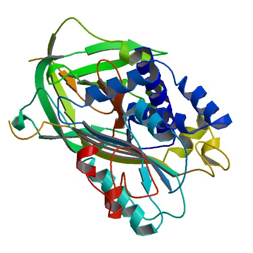
| + | [[Image:1oc0.jpg|frame|PAI-1 in complex with the SMB domain of vitronectin]] | ||
| + | |||
| + | Vitronectin is a glycoprotein found in the ECM and promotes cell adhesion and spreading. It has been shown to be implicated in hemostasis and tumor growth. Vitronectin regulates proteolysis initiated by plasminogen activation. | ||
| + | |||
| + | It interacts with the urokinase receptor through its Somatomedin B (SMB) domain, and this interaction has been implicated in cell migration and signal transduction. Vitronectin contains an Arginine, Glutamine, Aspartic acid (RGD) sequence that is a binding site for membrane bound integrins, which provides an anchor for cells to the ECM. High plasma levels of both Plasminogen Activator inhibitor-1 (PA-1)and the urokinase receptor have been shown to correlate with a negative prognosis for cancer patients. | ||
{{STRUCTURE_1oc0 | PDB=1oc0 | SCENE= }} | {{STRUCTURE_1oc0 | PDB=1oc0 | SCENE= }} | ||
| + | |||
| + | === Structure === | ||
| + | |||
| + | |||
| + | The spinning protein (<scene name='User:Yadilette Rivera-Colon/Sandbox_1/Loadedfrompdb/4'>Initial view</scene>) is the PAI-1 in complex with the SMB domain of vitronectin. | ||
| + | About one third of the Vitronectin's molecular mass is composed of carbohydrates. Occasionally the protein is cleaved after arginine 379, to produce two chain Vitronectin, where the two parts are linked by a disulfide bond. No high resolution structure has been determined experimentally yet. | ||
| + | |||
| + | Vitronectin consists of three domains: | ||
| + | |||
| + | * The N-terminal Somatomedin B domain (1-39). | ||
| + | * A central domains with hemopexin homology (131-342). | ||
| + | * A C-terminal domain (residues 347-459) also with hemopexin homology. | ||
Current revision
A CBI Molecule as a requirement for Chalk Talk offered in the University of Massachusetts Amherst Chemistry-Biology Interface Program at UMass Amherst and on display at the Molecular Playground.
Vitronectin
Vitronectin is a glycoprotein found in the ECM and promotes cell adhesion and spreading. It has been shown to be implicated in hemostasis and tumor growth. Vitronectin regulates proteolysis initiated by plasminogen activation.
It interacts with the urokinase receptor through its Somatomedin B (SMB) domain, and this interaction has been implicated in cell migration and signal transduction. Vitronectin contains an Arginine, Glutamine, Aspartic acid (RGD) sequence that is a binding site for membrane bound integrins, which provides an anchor for cells to the ECM. High plasma levels of both Plasminogen Activator inhibitor-1 (PA-1)and the urokinase receptor have been shown to correlate with a negative prognosis for cancer patients.
| |||||||||
| 1oc0, resolution 2.28Å () | |||||||||
|---|---|---|---|---|---|---|---|---|---|
| Related: | 1a7c, 1b3k, 1c5g, 1db2, 1dvm, 1dvn, 1lj5, 9pai | ||||||||
| |||||||||
| |||||||||
| Resources: | FirstGlance, OCA, RCSB, PDBsum | ||||||||
| Coordinates: | save as pdb, mmCIF, xml | ||||||||
Structure
The spinning protein () is the PAI-1 in complex with the SMB domain of vitronectin. About one third of the Vitronectin's molecular mass is composed of carbohydrates. Occasionally the protein is cleaved after arginine 379, to produce two chain Vitronectin, where the two parts are linked by a disulfide bond. No high resolution structure has been determined experimentally yet.
Vitronectin consists of three domains:
   * The N-terminal Somatomedin B domain (1-39).
   * A central domains with hemopexin homology (131-342).
   * A C-terminal domain (residues 347-459) also with hemopexin homology.


