This old version of Proteopedia is provided for student assignments while the new version is undergoing repairs. Content and edits done in this old version of Proteopedia after March 1, 2026 will eventually be lost when it is retired in about June of 2026.
Apply for new accounts at the new Proteopedia. Your logins will work in both the old and new versions.
Sandbox Reserved 14
From Proteopedia
(Difference between revisions)
| (50 intermediate revisions not shown.) | |||
| Line 1: | Line 1: | ||
| - | + | <!-- DO NOT DELETE THE TEMPLATE LINE --> | |
| + | {{Template:Sandbox Reserved Eric Martz}} | ||
| + | <!-- INSERT YOUR SCENES AND TEXT BELOW THIS LINE --> | ||
| - | == | + | <Structure load='1d66' size='350' frame='true' align='right' caption='Insert caption here' scene='Insert optional scene name here' /> |
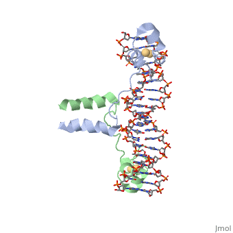
| - | + | This is the DNA-binding domain of the gal4 transcriptional regulatory protein, bound to DNA. | |
| + | *<scene name='42/421644/Osaka1/1'>Here the DNA is solid</scene> | ||
| + | (atoms have van der Waals radii). | ||
| + | *The DNA-binding surface of the protein has <scene name='42/421644/Osaka1/2'>only positive charges</scene> to bind to the negatively charged DNA phosphate backbone. | ||
| + | (<font color="blue">Positive</font>, | ||
| + | <font color="red">Negative</font>) | ||
Current revision
| This Sandbox is Reserved from May 10, 2015, through July 31, 2015 for use by the class Protein 3D Structure Visualization & Structural Bioinformatics taught by Eric Martz and Keiichi Namba at Osaka University, Japan. This reservation includes Sandbox Reserved 1 through Sandbox Reserved 10. Syllabus. |
To get started:
More help: Getting Started in Proteopedia, Help:Editing, Main Help Page. |
|
This is the DNA-binding domain of the gal4 transcriptional regulatory protein, bound to DNA.
(atoms have van der Waals radii).
- The DNA-binding surface of the protein has to bind to the negatively charged DNA phosphate backbone.
(Positive, Negative)
