This old version of Proteopedia is provided for student assignments while the new version is undergoing repairs. Content and edits done in this old version of Proteopedia after March 1, 2026 will eventually be lost when it is retired in about June of 2026.
Apply for new accounts at the new Proteopedia. Your logins will work in both the old and new versions.
2avi
From Proteopedia
(Difference between revisions)
m (Protected "2avi" [edit=sysop:move=sysop]) |
|||
| (9 intermediate revisions not shown.) | |||
| Line 1: | Line 1: | ||
| - | [[Image:2avi.png|left|200px]] | ||
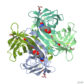
| - | < | + | ==THREE-DIMENSIONAL STRUCTURES OF AVIDIN AND THE AVIDIN-BIOTIN COMPLEX== |
| - | + | <StructureSection load='2avi' size='340' side='right'caption='[[2avi]], [[Resolution|resolution]] 3.00Å' scene=''> | |
| - | You may | + | == Structural highlights == |
| - | + | <table><tr><td colspan='2'>[[2avi]] is a 2 chain structure with sequence from [https://en.wikipedia.org/wiki/Gallus_gallus Gallus gallus]. Full crystallographic information is available from [http://oca.weizmann.ac.il/oca-bin/ocashort?id=2AVI OCA]. For a <b>guided tour on the structure components</b> use [https://proteopedia.org/fgij/fg.htm?mol=2AVI FirstGlance]. <br> | |
| - | + | </td></tr><tr id='method'><td class="sblockLbl"><b>[[Empirical_models|Method:]]</b></td><td class="sblockDat" id="methodDat">X-ray diffraction, [[Resolution|Resolution]] 3Å</td></tr> | |
| - | - | + | <tr id='ligand'><td class="sblockLbl"><b>[[Ligand|Ligands:]]</b></td><td class="sblockDat" id="ligandDat"><scene name='pdbligand=BTN:BIOTIN'>BTN</scene>, <scene name='pdbligand=NAG:N-ACETYL-D-GLUCOSAMINE'>NAG</scene></td></tr> |
| - | + | <tr id='resources'><td class="sblockLbl"><b>Resources:</b></td><td class="sblockDat"><span class='plainlinks'>[https://proteopedia.org/fgij/fg.htm?mol=2avi FirstGlance], [http://oca.weizmann.ac.il/oca-bin/ocaids?id=2avi OCA], [https://pdbe.org/2avi PDBe], [https://www.rcsb.org/pdb/explore.do?structureId=2avi RCSB], [https://www.ebi.ac.uk/pdbsum/2avi PDBsum], [https://prosat.h-its.org/prosat/prosatexe?pdbcode=2avi ProSAT]</span></td></tr> | |
| + | </table> | ||
| + | == Function == | ||
| + | [https://www.uniprot.org/uniprot/AVID_CHICK AVID_CHICK] The biological function of avidin is not known. Forms a strong non-covalent specific complex with biotin (one molecule of biotin per subunit of avidin). | ||
| + | == Evolutionary Conservation == | ||
| + | [[Image:Consurf_key_small.gif|200px|right]] | ||
| + | Check<jmol> | ||
| + | <jmolCheckbox> | ||
| + | <scriptWhenChecked>; select protein; define ~consurf_to_do selected; consurf_initial_scene = true; script "/wiki/ConSurf/av/2avi_consurf.spt"</scriptWhenChecked> | ||
| + | <scriptWhenUnchecked>script /wiki/extensions/Proteopedia/spt/initialview01.spt</scriptWhenUnchecked> | ||
| + | <text>to colour the structure by Evolutionary Conservation</text> | ||
| + | </jmolCheckbox> | ||
| + | </jmol>, as determined by [http://consurfdb.tau.ac.il/ ConSurfDB]. You may read the [[Conservation%2C_Evolutionary|explanation]] of the method and the full data available from [http://bental.tau.ac.il/new_ConSurfDB/main_output.php?pdb_ID=2avi ConSurf]. | ||
| + | <div style="clear:both"></div> | ||
| + | <div style="background-color:#fffaf0;"> | ||
| + | == Publication Abstract from PubMed == | ||
| + | The crystal structures of a deglycosylated form of the egg-white glycoprotein avidin and of its complex with biotin have been determined to 2.6 and 3.0 A, respectively. The structures reveal the amino acid residues critical for stabilization of the tetrameric assembly and for the exceptionally tight binding of biotin. Each monomer is an eight-stranded antiparallel beta-barrel, remarkably similar to that of the genetically distinct bacterial analog streptavidin. As in streptavidin, binding of biotin involves a highly stabilized network of polar and hydrophobic interactions. There are, however, some differences. The presence of additional hydrophobic and hydrophilic groups in the binding site of avidin (which are missing in streptavidin) may account for its higher affinity constant. Two amino acid substitutions are proposed to be responsible for its susceptibility to denaturation relative to streptavidin. Unexpectedly, a residual N-acetylglucosamine moiety was detected in the deglycosylated avidin monomer by difference Fourier synthesis. | ||
| - | + | Three-dimensional structures of avidin and the avidin-biotin complex.,Livnah O, Bayer EA, Wilchek M, Sussman JL Proc Natl Acad Sci U S A. 1993 Jun 1;90(11):5076-80. PMID:8506353<ref>PMID:8506353</ref> | |
| - | + | From MEDLINE®/PubMed®, a database of the U.S. National Library of Medicine.<br> | |
| - | + | </div> | |
| - | + | <div class="pdbe-citations 2avi" style="background-color:#fffaf0;"></div> | |
| - | + | ||
| - | + | ||
| - | + | ||
| - | + | ||
| - | == | + | |
| - | + | ||
==See Also== | ==See Also== | ||
| - | *[[Avidin]] | + | *[[Avidin|Avidin]] |
| - | + | *[[Avidin 3D structures|Avidin 3D structures]] | |
| - | == | + | == References == |
| - | < | + | <references/> |
| + | __TOC__ | ||
| + | </StructureSection> | ||
[[Category: Gallus gallus]] | [[Category: Gallus gallus]] | ||
| - | [[Category: | + | [[Category: Large Structures]] |
| - | [[Category: | + | [[Category: Livnah O]] |
| - | [[Category: | + | [[Category: Sussman J]] |
Current revision
THREE-DIMENSIONAL STRUCTURES OF AVIDIN AND THE AVIDIN-BIOTIN COMPLEX
| |||||||||||