This old version of Proteopedia is provided for student assignments while the new version is undergoing repairs. Content and edits done in this old version of Proteopedia after March 1, 2026 will eventually be lost when it is retired in about June of 2026.
Apply for new accounts at the new Proteopedia. Your logins will work in both the old and new versions.
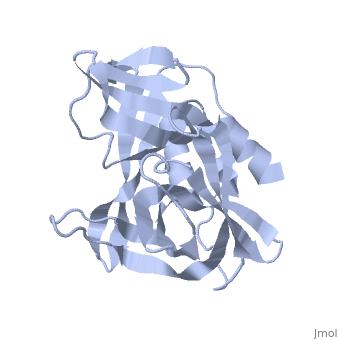
2j92
From Proteopedia
(Difference between revisions)
| (9 intermediate revisions not shown.) | |||
| Line 1: | Line 1: | ||
| - | [[Image:2j92.png|left|200px]] | ||
| - | + | ==3C PROTEASE FROM TYPE A10(61) FOOT-AND-MOUTH DISEASE VIRUS - Crystal packing mutant (K51Q)== | |
| - | + | <StructureSection load='2j92' size='340' side='right'caption='[[2j92]], [[Resolution|resolution]] 2.20Å' scene=''> | |
| - | + | == Structural highlights == | |
| - | + | <table><tr><td colspan='2'>[[2j92]] is a 2 chain structure with sequence from [https://en.wikipedia.org/wiki/Foot-and-mouth_disease_virus_(strain_A10-61) Foot-and-mouth disease virus (strain A10-61)]. Full crystallographic information is available from [http://oca.weizmann.ac.il/oca-bin/ocashort?id=2J92 OCA]. For a <b>guided tour on the structure components</b> use [https://proteopedia.org/fgij/fg.htm?mol=2J92 FirstGlance]. <br> | |
| - | + | </td></tr><tr id='method'><td class="sblockLbl"><b>[[Empirical_models|Method:]]</b></td><td class="sblockDat" id="methodDat">X-ray diffraction, [[Resolution|Resolution]] 2.2Å</td></tr> | |
| - | --> | + | <tr id='resources'><td class="sblockLbl"><b>Resources:</b></td><td class="sblockDat"><span class='plainlinks'>[https://proteopedia.org/fgij/fg.htm?mol=2j92 FirstGlance], [http://oca.weizmann.ac.il/oca-bin/ocaids?id=2j92 OCA], [https://pdbe.org/2j92 PDBe], [https://www.rcsb.org/pdb/explore.do?structureId=2j92 RCSB], [https://www.ebi.ac.uk/pdbsum/2j92 PDBsum], [https://prosat.h-its.org/prosat/prosatexe?pdbcode=2j92 ProSAT]</span></td></tr> |
| - | + | </table> | |
| + | == Function == | ||
| + | [https://www.uniprot.org/uniprot/POLG_FMDV1 POLG_FMDV1] The leader protease autocatalytically cleaves itself from the polyprotein at the L/VP0 junction. It cleaves the host translation initiation factors EIF4G1 and EIF4G3, in order to shut down the capped cellular mRNA transcription (By similarity). Capsid proteins VP1, VP2, VP3 and VP4 form a closed capsid enclosing the viral positive strand RNA genome. VP4 lies on the inner surface of the protein shell formed by VP1, VP2 and VP3. All the three latter proteins contain a beta-sheet structure called beta-barrel jelly roll. Together they form an icosahedral capsid (T=3) composed of 60 copies of each VP1, VP2, and VP3, with a diameter of approximately 300 Angstroms. VP1 is situated at the 12 fivefold axes, whereas VP2 and VP3 are located at the quasi-sixfold axes. The capsid interacts with host heparan sulfate and various integrins (alphavbeta1, alphavbeta3, alpha5beta1, alphavbeta6, alphavbeta8) to provide virion attachment to target Attachment via host integrins induces virion internalization predominantly through clathrin-mediated endocytosis (By similarity). Protein VP0: VP0 precursor is a component of immature procapsids (By similarity). Protein 2B: Affects membrane integrity and cause an increase in membrane permeability (By similarity). Protein 2C: Associates with and induces structural rearrangements of intracellular membranes. It displays RNA-binding, nucleotide binding and NTPase activities (By similarity). Protein 3A, via its hydrophobic domain, serves as membrane anchor (By similarity). Protein 3B-1, 3B-2 and 3B-3 are covalently linked to the 5'-end of both the positive-strand and negative-strand genomic RNAs. They acts as a genome-linked replication primer (By similarity). Protease 3C: cysteine protease that generates mature viral proteins from the precursor polyprotein. In addition to its proteolytic activity, it binds to viral RNA, and thus influences viral genome replication. RNA and substrate bind cooperatively to the protease (By similarity). RNA-directed RNA polymerase 3D-POL replicates genomic and antigenomic RNA by recognizing replications specific signals (By similarity). | ||
| + | == Evolutionary Conservation == | ||
| + | [[Image:Consurf_key_small.gif|200px|right]] | ||
| + | Check<jmol> | ||
| + | <jmolCheckbox> | ||
| + | <scriptWhenChecked>; select protein; define ~consurf_to_do selected; consurf_initial_scene = true; script "/wiki/ConSurf/j9/2j92_consurf.spt"</scriptWhenChecked> | ||
| + | <scriptWhenUnchecked>script /wiki/extensions/Proteopedia/spt/initialview01.spt</scriptWhenUnchecked> | ||
| + | <text>to colour the structure by Evolutionary Conservation</text> | ||
| + | </jmolCheckbox> | ||
| + | </jmol>, as determined by [http://consurfdb.tau.ac.il/ ConSurfDB]. You may read the [[Conservation%2C_Evolutionary|explanation]] of the method and the full data available from [http://bental.tau.ac.il/new_ConSurfDB/main_output.php?pdb_ID=2j92 ConSurf]. | ||
| + | <div style="clear:both"></div> | ||
| + | <div style="background-color:#fffaf0;"> | ||
| + | == Publication Abstract from PubMed == | ||
| + | The 3C protease (3C(pro)) from foot-and-mouth disease virus (FMDV), the causative agent of a widespread and economically devastating disease of domestic livestock, is a potential target for antiviral drug design. We have determined the structure of a new crystal form of FMDV 3C(pro), a chymotrypsin-like cysteine protease, which reveals features that are important for catalytic activity. In particular, we show that a surface loop which was disordered in previous structures adopts a beta-ribbon structure that is conformationally similar to equivalent regions on other picornaviral 3C proteases and some serine proteases. This beta-ribbon folds over the peptide binding cleft and clearly contributes to substrate recognition. Replacement of Cys142 at the tip of the beta-ribbon with different amino acids has a significant impact on enzyme activity and shows that higher activity is obtained with more hydrophobic side chains. Comparison of the structure of FMDV 3C(pro) with homologous enzyme-peptide complexes suggests that this correlation arises because the side chain of Cys142 contacts the hydrophobic portions of the P2 and P4 residues in the peptide substrate. Collectively, these findings provide compelling evidence for the role of the beta-ribbon in catalytic activity and provide valuable insights for the design of FMDV 3C(pro) inhibitors. | ||
| - | + | Structural and mutagenic analysis of foot-and-mouth disease virus 3C protease reveals the role of the beta-ribbon in proteolysis.,Sweeney TR, Roque-Rosell N, Birtley JR, Leatherbarrow RJ, Curry S J Virol. 2007 Jan;81(1):115-24. Epub 2006 Oct 25. PMID:17065215<ref>PMID:17065215</ref> | |
| + | From MEDLINE®/PubMed®, a database of the U.S. National Library of Medicine.<br> | ||
| + | </div> | ||
| + | <div class="pdbe-citations 2j92" style="background-color:#fffaf0;"></div> | ||
| - | + | ==See Also== | |
| - | + | *[[Virus protease 3D structures|Virus protease 3D structures]] | |
| - | + | == References == | |
| - | + | <references/> | |
| - | + | __TOC__ | |
| - | + | </StructureSection> | |
| - | == | + | [[Category: Large Structures]] |
| - | [[ | + | [[Category: Birtley JR]] |
| - | + | [[Category: Curry S]] | |
| - | == | + | [[Category: Leatherbarrow RJ]] |
| - | < | + | [[Category: Sweeney TR]] |
| - | [[Category: | + | |
| - | + | ||
| - | [[Category: Birtley | + | |
| - | [[Category: Curry | + | |
| - | [[Category: Leatherbarrow | + | |
| - | [[Category: Sweeney | + | |
| - | + | ||
| - | + | ||
| - | + | ||
| - | + | ||
| - | + | ||
Current revision
3C PROTEASE FROM TYPE A10(61) FOOT-AND-MOUTH DISEASE VIRUS - Crystal packing mutant (K51Q)
| |||||||||||