The Structure and Mechanism of Hexokinase
From Proteopedia
(Difference between revisions)
| (8 intermediate revisions not shown.) | |||
| Line 1: | Line 1: | ||
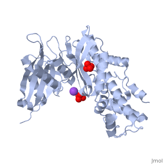
| - | < | + | <StructureSection load='1v4t' size='400' side='right' scene='' caption='Structure of human Glucokinase complex with sulfate and Na+ ion [[1v4t]]'> |
| - | A hexokinase is an enzyme that phosphorylates a six-carbon sugar, a hexose, to a hexose phosphate. In most tissues and organisms, glucose is the most important substrate of hexokinases, and glucose 6-phosphate the most important product. Hexokinases have been found in every organism checked, ranging from bacteria, yeast, and plants, to humans and other vertebrates. They are categorized as actin fold proteins, sharing a common ATP binding site core surrounded by more variable sequences that determine substrate affinities and other properties. Several hexokinase isoforms or isozymes providing different functions can occur in a single species. | + | |
| + | A [[hexokinase]] is an enzyme that phosphorylates a six-carbon sugar, a hexose, to a hexose phosphate. In most tissues and organisms, glucose is the most important substrate of hexokinases, and glucose 6-phosphate the most important product. Hexokinases have been found in every organism checked, ranging from bacteria, yeast, and plants, to humans and other vertebrates. They are categorized as actin fold proteins, sharing a common ATP binding site core surrounded by more variable sequences that determine substrate affinities and other properties. Several hexokinase isoforms or isozymes providing different functions can occur in a single species. | ||
-Hexokinase I/A is found in all mammalian tissues, and is considered a "housekeeping enzyme," unaffected by most physiological, hormonal, and metabolic changes. | -Hexokinase I/A is found in all mammalian tissues, and is considered a "housekeeping enzyme," unaffected by most physiological, hormonal, and metabolic changes. | ||
| Line 31: | Line 32: | ||
'''Glucokinase vs. Other Hexokinases:''' Glucokinase is unique from other hexokinase in kinetic properties and is coded by a different gene. The difference of glucokinase from the other hexokinases is that glucokinase has a lower affinity, thus a higher Km, for glucose. The reduced affinity for glucose allows the activity of glucokinase to differ under physiological conditions according to the amount of glucose present. Essentially, this means that it operates only when serum glucose levels are high. Other tissues need to use glucose at lower serum levels and thus use the higher affinity (lower Km) hexokinase. Glucose-6-phosphate inhibits hexokinase and, if the cell is not using up the G6P that it is making, then it should stop making it, thus making the process a product inhibition. In contrast, G6P does not inhibit glucokinase, and this allows it to remain active in storing as much glucose as possible in the presence of high glucose levels. | '''Glucokinase vs. Other Hexokinases:''' Glucokinase is unique from other hexokinase in kinetic properties and is coded by a different gene. The difference of glucokinase from the other hexokinases is that glucokinase has a lower affinity, thus a higher Km, for glucose. The reduced affinity for glucose allows the activity of glucokinase to differ under physiological conditions according to the amount of glucose present. Essentially, this means that it operates only when serum glucose levels are high. Other tissues need to use glucose at lower serum levels and thus use the higher affinity (lower Km) hexokinase. Glucose-6-phosphate inhibits hexokinase and, if the cell is not using up the G6P that it is making, then it should stop making it, thus making the process a product inhibition. In contrast, G6P does not inhibit glucokinase, and this allows it to remain active in storing as much glucose as possible in the presence of high glucose levels. | ||
| - | <applet load='1v4t' size='300' color='white' frame='true' align='right' caption='Structure of Glucokinase' /> | ||
'''Glucokinase Structure:''' Glucokinase also contains <scene name='Kyle_Schroering_Sandbox/Open_conformation/1'>inactive</scene> and <scene name='Kyle_Schroering_Sandbox/Closed_conformation/1'>active</scene> conformation. Glucokinase consists of one chain or subunit of 448 amino acids forming a monomeric molecule consisting of 13 alpha helices and 5 beta sheets that can phosporylate glucose and other hexoses. The chain is folded into two distinct regions, a small and large domain. Glucokinase has one active binding site for glucose and one for ATP, which is the energy source for phosphorylation. This active binding site is located between the small and large domains. The carboxyl terminus is part of the alpha 13 helix, which codes for the region that forms half of the binding site for glucose. Glucokinase can be modulated to form an inactive and active complex. The inactive conformation forms when the alpha 13 helix has been modulated away from the rest of the molecule forming a large space. This space is too large to bind glucose so it is said to be in the inactive form. The alternative is when the alpha 13 helix is modulated to form a smaller space thus activating the protein<ref name="king">PMID:15016359</ref>. | '''Glucokinase Structure:''' Glucokinase also contains <scene name='Kyle_Schroering_Sandbox/Open_conformation/1'>inactive</scene> and <scene name='Kyle_Schroering_Sandbox/Closed_conformation/1'>active</scene> conformation. Glucokinase consists of one chain or subunit of 448 amino acids forming a monomeric molecule consisting of 13 alpha helices and 5 beta sheets that can phosporylate glucose and other hexoses. The chain is folded into two distinct regions, a small and large domain. Glucokinase has one active binding site for glucose and one for ATP, which is the energy source for phosphorylation. This active binding site is located between the small and large domains. The carboxyl terminus is part of the alpha 13 helix, which codes for the region that forms half of the binding site for glucose. Glucokinase can be modulated to form an inactive and active complex. The inactive conformation forms when the alpha 13 helix has been modulated away from the rest of the molecule forming a large space. This space is too large to bind glucose so it is said to be in the inactive form. The alternative is when the alpha 13 helix is modulated to form a smaller space thus activating the protein<ref name="king">PMID:15016359</ref>. | ||
| - | Glucokinase includes the < | + | Glucokinase includes the <jmol><jmolLink><script>script "/scripts/Kyle_Schroering_Sandbox/1v4s_glucose/2.spt"; select not *.CB;label off</script><text>glucose binding site (active form)</text></jmolLink></jmol> where glucose forms hydrogen bonds at the bottom of the deep crevice between the large domain and the small domain. E256, E290 (shown in green) of the large domain, T168, K169 (shown in red) of the small domain, and N204, D205 (shown in yellow) of a connecting region form hydrogen bonds with glucose. The <jmol><jmolLink><script>script "/scripts/Kyle_Schroering_Sandbox/Active_form_of_glucokinase/4.spt"; select not *.CB;label off</script><text>glucose binding site (inactive form)</text></jmolLink></jmol> shows a different conformation. At the <jmol><jmolLink><script>script "/scripts/Kyle_Schroering_Sandbox/1v4s_atp/3.spt"; select not *.CB;label off</script><text>allosteric site (active form)</text></jmolLink></jmol>, ATP forms hydrogen bonds with R63 and Y215 (shown in orange) and hydrophobically interacts with M210, Y214 (shown in blue) of the α5 helix and V452, V455 (shown in green) of the α13 helix. The <scene name='38/387916/Allosteric/1'>allosteric site (inactive form)</scene> again shows structural differences. The differences in these two conformations allows glucokinase to function properly in different levels of glucose concentration. |
'''Proposed Mechanism for Glucokinase:''' As described above, glucokinase has a distinct conformation change from the active and inactive form. Experiments have also shown an intermediate open form based on analysis of the movement between the active and inactive form. The switch in conformations between the active form and the intermediate is a kinetically faster step than the change between the intermediate and the inactive form. The inactive form of gluckokinase is the thermodynamically favored unless there is glucose present. Glucokinase does not change conformation until the glucose molecule binds. The conformation change may be triggered by the interaction between Asp 205 and the glucose molecule. Once glucokinase is in the active form, the enzymatic reaction is carried out with the presence of ATP. All in all, glucose binds to glucokinase and then is phosphorylated by ATP to produce glucose-6-phosphate and ADP.<ref name="king">PMID:15016359</ref> | '''Proposed Mechanism for Glucokinase:''' As described above, glucokinase has a distinct conformation change from the active and inactive form. Experiments have also shown an intermediate open form based on analysis of the movement between the active and inactive form. The switch in conformations between the active form and the intermediate is a kinetically faster step than the change between the intermediate and the inactive form. The inactive form of gluckokinase is the thermodynamically favored unless there is glucose present. Glucokinase does not change conformation until the glucose molecule binds. The conformation change may be triggered by the interaction between Asp 205 and the glucose molecule. Once glucokinase is in the active form, the enzymatic reaction is carried out with the presence of ATP. All in all, glucose binds to glucokinase and then is phosphorylated by ATP to produce glucose-6-phosphate and ADP.<ref name="king">PMID:15016359</ref> | ||
| Line 45: | Line 45: | ||
In the pancreas, a rise in glucose levels increases the activity of glucokinase causing an increase in glucose 6-phosphate. This causes the triggering of the beta cells to secret insulin<ref>PMID:11237213</ref>. Glucokinase is the first step in this reaction. Insulin then allows other cells in the body to take up glucose, actively lowering the glucose level | In the pancreas, a rise in glucose levels increases the activity of glucokinase causing an increase in glucose 6-phosphate. This causes the triggering of the beta cells to secret insulin<ref>PMID:11237213</ref>. Glucokinase is the first step in this reaction. Insulin then allows other cells in the body to take up glucose, actively lowering the glucose level | ||
| + | </StructureSection> | ||
| + | ==3D structures of hexokinase== | ||
| - | + | [[Hexokinase]] | |
Current revision
| |||||||||||
3D structures of hexokinase
Additional Resources
For additional information, see: Carbohydrate Metabolism
References
- ↑ Pollard-Knight D, Cornish-Bowden A. Mechanism of liver glucokinase. Mol Cell Biochem. 1982 Apr 30;44(2):71-80. PMID:7048063
- ↑ Koshland D. Induced Fit and Hexokinase.Fig.7 Active Site of Glucokinase/Hexokinase.2010; [1]
- ↑ Kitto B.G, Caras J., Caras P. Interactive Concepts in Biochemistry. Structural Tutorials.2008; Chp: 15.1, 15.5. [ http://higheredbcs.wiley.com/legacy/college/boyer/0471661791/structure/hexokinase/hexokinase.htm]
- ↑ 4.0 4.1 4.2 4.3 4.4 Kamata K, Mitsuya M, Nishimura T, Eiki J, Nagata Y. Structural basis for allosteric regulation of the monomeric allosteric enzyme human glucokinase. Structure. 2004 Mar;12(3):429-38. PMID:15016359 doi:10.1016/j.str.2004.02.005
- ↑ Postic C, Shiota M, Magnuson MA. Cell-specific roles of glucokinase in glucose homeostasis. Recent Prog Horm Res. 2001;56:195-217. PMID:11237213
Proteopedia Page Contributors and Editors (what is this?)
Kyle Schroering, Ann Taylor, Michal Harel, Drew McCaffrey, Alexander Berchansky, Karsten Theis, Joel L. Sussman, Cody Leatherman, David Canner
