This old version of Proteopedia is provided for student assignments while the new version is undergoing repairs. Content and edits done in this old version of Proteopedia after March 1, 2026 will eventually be lost when it is retired in about June of 2026.
Apply for new accounts at the new Proteopedia. Your logins will work in both the old and new versions.
Parvalbumin
From Proteopedia
(Difference between revisions)
(New page: Crystal Structure of Parvalbumin 4cpv {{STRUCTURE_4cpv| PDB=4cpv | SIZE=300| SCENE= |right|CAPTION=Parvalbumin 4cpv }}) |
|||
| (30 intermediate revisions not shown.) | |||
| Line 1: | Line 1: | ||
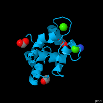
| - | [[ | + | <StructureSection load='' size='350' side='right' caption='Pike parvalbumin complex with Ca+2 ions (green), ammonium ion (blue), acetyl and formate (PDB entry [[2pvb]])' scene='46/466520/Cv/2' pspeed='8'> |
| - | {{ | + | == Function == |
| + | '''Parvalbumin (PVA)''' is a calcium-binding albumin protein. PVA is involved in calcium signaling<ref>PMID:11867433</ref>. PVA contains 3 domains each containing 2 helices: AB, CD (N-terminal) and EF (C-terminal). '''α-parvalbumin''' differs from '''β-parvalbumin''' in 54 positions. α-parvalbumin is expressed in various tissues while β-parvalbumin is expressed only in preterm placenta<ref>PMID:8354278</ref>. <br /> | ||
| + | See also<br /> | ||
| + | * [[EF hand]] | ||
| + | * [[Parvalbumin carp]]. | ||
| + | |||
| + | == Relevance == | ||
| + | PVA is a major fish allergen<ref>PMID:18221468</ref>. | ||
| + | |||
| + | == Structural highlights == | ||
| + | PVA contains a <scene name='46/466520/Cv/10'>heptacoordinated Ca+2 ion binding to 7 oxygen atoms</scene> in a <scene name='46/466520/Cv/11'>typical EF-hand</scene> coordination<ref>PMID:10548066</ref>. Water molecules are shown as red spheres. <scene name='46/466520/Cv/12'>Calcium ion is coordinated by seven oxygen atoms that form a pentagonal bipyramid </scene>. | ||
| + | *<scene name='46/466520/Cv/13'>Ammonium ion coordination site</scene>. | ||
| + | </StructureSection> | ||
| + | == 3D Structures of Parvalbumin == | ||
| + | Updated on {{REVISIONDAY2}}-{{MONTHNAME|{{REVISIONMONTH}}}}-{{REVISIONYEAR}} | ||
| + | {{#tree:id=OrganizedByTopic|openlevels=0| | ||
| + | |||
| + | *parvalbumin | ||
| + | |||
| + | **[[4cpv]], [[5cpv]] – cPVA - carp<br /> | ||
| + | **[[1cdp]] – cPVA Cd-substituted<br /> | ||
| + | **[[1b8c]], [[1b8r]], [[1b9a]], [[1b8l]] – cPVA (mutant)<br /> | ||
| + | **[[1pal]], [[2pal]], [[3pal]], [[4pal]], [[1pvb]], [[1pva]], [[2pvb]] – pPVA – pike<br /> | ||
| + | **[[2pas]], [[3pat]] – pPVA – NMR<br /> | ||
| + | **[[1a75]] – PVA – whiting<br /> | ||
| + | **[[1bu3]] – PVA Ca-binding domain – hake<br /> | ||
| + | **[[5zgm]] – MgPVA SPV-I – ''Mustelus griseus''<br /> | ||
| + | **[[5zh6]] – MgPVA SPV-II<br /> | ||
| + | |||
| + | *α-parvalbumin | ||
| + | |||
| + | **[[1rjv]], [[1rk9]] - hPVA-α - human – NMR<br /> | ||
| + | **[[1rtp]], [[1rwy]] - rPVA-α - rat<br /> | ||
| + | **[[2jww]] - rPVA-α Ca-free – NMR<br /> | ||
| + | **[[1g33]], [[1s3p]], [[1xvj]], [[3f45]] - rPVA-α (mutant)<br /> | ||
| + | **[[5pal]] – PVA-α - shark<br /> | ||
| + | |||
| + | *β-parvalbumin (oncomodulin) | ||
| + | |||
| + | **[[1ttx]] - hPVA-β (mutant) – NMR<br /> | ||
| + | **[[2nln]] - rPVA-β Ca-free – NMR<br /> | ||
| + | **[[1rro]], [[1omd]] - rPVA-β <br /> | ||
| + | **[[3fs7]] - chPVA-β - chicken<br /> | ||
| + | **[[2kqy]] – chPVA-β – NMR<br /> | ||
| + | **[[2kyc]] - chPVA-β <br /> | ||
| + | **[[2kyf]] - chPVA-β (mutant) – NMR<br /> | ||
| + | **[[2mbx]] - PVA-β – cod - NMR<br /> | ||
| + | **[[5xnd]] - PVA-β – mackerel - NMR<br /> | ||
| + | }} | ||
| + | == References == | ||
| + | <references/> | ||
| + | [[Category:Topic Page]] | ||
Current revision
| |||||||||||
3D Structures of Parvalbumin
Updated on 05-December-2021
References
- ↑ Cates MS, Teodoro ML, Phillips GN Jr. Molecular mechanisms of calcium and magnesium binding to parvalbumin. Biophys J. 2002 Mar;82(3):1133-46. PMID:11867433 doi:http://dx.doi.org/10.1016/S0006-3495(02)75472-6
- ↑ Fohr UG, Weber BR, Muntener M, Staudenmann W, Hughes GJ, Frutiger S, Banville D, Schafer BW, Heizmann CW. Human alpha and beta parvalbumins. Structure and tissue-specific expression. Eur J Biochem. 1993 Aug 1;215(3):719-27. PMID:8354278
- ↑ Lim DL, Neo KH, Yi FC, Chua KY, Goh DL, Shek LP, Giam YC, Van Bever HP, Lee BW. Parvalbumin--the major tropical fish allergen. Pediatr Allergy Immunol. 2008 Aug;19(5):399-407. doi:, 10.1111/j.1399-3038.2007.00674.x. Epub 2008 Jan 25. PMID:18221468 doi:http://dx.doi.org/10.1111/j.1399-3038.2007.00674.x
- ↑ Declercq JP, Evrard C, Lamzin V, Parello J. Crystal structure of the EF-hand parvalbumin at atomic resolution (0.91 A) and at low temperature (100 K). Evidence for conformational multistates within the hydrophobic core. Protein Sci. 1999 Oct;8(10):2194-204. PMID:10548066
