This old version of Proteopedia is provided for student assignments while the new version is undergoing repairs. Content and edits done in this old version of Proteopedia after March 1, 2026 will eventually be lost when it is retired in about June of 2026.
Apply for new accounts at the new Proteopedia. Your logins will work in both the old and new versions.
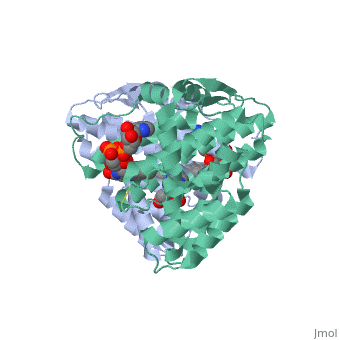
3p0k
From Proteopedia
(Difference between revisions)
| (5 intermediate revisions not shown.) | |||
| Line 1: | Line 1: | ||
| - | [[Image:3p0k.jpg|left|200px]] | ||
| - | < | + | ==Structure of Baculovirus Sulfhydryl Oxidase Ac92== |
| - | + | <StructureSection load='3p0k' size='340' side='right'caption='[[3p0k]], [[Resolution|resolution]] 1.47Å' scene=''> | |
| - | + | == Structural highlights == | |
| - | + | <table><tr><td colspan='2'>[[3p0k]] is a 1 chain structure with sequence from [https://en.wikipedia.org/wiki/Autographa_californica_nucleopolyhedrovirus Autographa californica nucleopolyhedrovirus]. Full crystallographic information is available from [http://oca.weizmann.ac.il/oca-bin/ocashort?id=3P0K OCA]. For a <b>guided tour on the structure components</b> use [https://proteopedia.org/fgij/fg.htm?mol=3P0K FirstGlance]. <br> | |
| - | + | </td></tr><tr id='method'><td class="sblockLbl"><b>[[Empirical_models|Method:]]</b></td><td class="sblockDat" id="methodDat">X-ray diffraction, [[Resolution|Resolution]] 1.47Å</td></tr> | |
| - | --> | + | <tr id='ligand'><td class="sblockLbl"><b>[[Ligand|Ligands:]]</b></td><td class="sblockDat" id="ligandDat"><scene name='pdbligand=ACT:ACETATE+ION'>ACT</scene>, <scene name='pdbligand=FAD:FLAVIN-ADENINE+DINUCLEOTIDE'>FAD</scene>, <scene name='pdbligand=IMD:IMIDAZOLE'>IMD</scene></td></tr> |
| - | + | <tr id='resources'><td class="sblockLbl"><b>Resources:</b></td><td class="sblockDat"><span class='plainlinks'>[https://proteopedia.org/fgij/fg.htm?mol=3p0k FirstGlance], [http://oca.weizmann.ac.il/oca-bin/ocaids?id=3p0k OCA], [https://pdbe.org/3p0k PDBe], [https://www.rcsb.org/pdb/explore.do?structureId=3p0k RCSB], [https://www.ebi.ac.uk/pdbsum/3p0k PDBsum], [https://prosat.h-its.org/prosat/prosatexe?pdbcode=3p0k ProSAT]</span></td></tr> | |
| + | </table> | ||
| + | == Function == | ||
| + | [https://www.uniprot.org/uniprot/FLSO_NPVAC FLSO_NPVAC] Functional FAD-linked sulfhydryl oxidase that is required for infectious budded virion (BV) production and for the formation of enveloped occluded virion (ODV).<ref>PMID:19409596</ref> | ||
| - | == | + | ==See Also== |
| - | + | *[[Sulfhydryl oxidase|Sulfhydryl oxidase]] | |
| - | + | *[[Sulfhydryl oxidase 3D structures|Sulfhydryl oxidase 3D structures]] | |
| - | + | == References == | |
| - | + | <references/> | |
| - | + | __TOC__ | |
| - | + | </StructureSection> | |
| - | + | ||
| - | + | ||
| - | + | ||
| - | [[ | + | |
| - | + | ||
| - | == | + | |
| - | < | + | |
[[Category: Autographa californica nucleopolyhedrovirus]] | [[Category: Autographa californica nucleopolyhedrovirus]] | ||
| - | [[Category: | + | [[Category: Large Structures]] |
| - | [[Category: Fass | + | [[Category: Fass D]] |
| - | [[Category: Hakim | + | [[Category: Hakim M]] |
| - | + | ||
| - | + | ||
| - | + | ||
| - | + | ||
| - | + | ||
| - | + | ||
Current revision
Structure of Baculovirus Sulfhydryl Oxidase Ac92
| |||||||||||