1gm5
From Proteopedia
(Difference between revisions)
m (Protected "1gm5" [edit=sysop:move=sysop]) |
|||
| (10 intermediate revisions not shown.) | |||
| Line 1: | Line 1: | ||
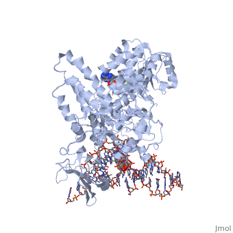
| - | [[Image:1gm5.png|left|200px]] | ||
| - | + | ==Structure of RecG bound to three-way DNA junction== | |
| - | + | <StructureSection load='1gm5' size='340' side='right'caption='[[1gm5]], [[Resolution|resolution]] 3.24Å' scene=''> | |
| - | + | == Structural highlights == | |
| - | + | <table><tr><td colspan='2'>[[1gm5]] is a 4 chain structure with sequence from [https://en.wikipedia.org/wiki/Thermotoga_maritima Thermotoga maritima]. Full crystallographic information is available from [http://oca.weizmann.ac.il/oca-bin/ocashort?id=1GM5 OCA]. For a <b>guided tour on the structure components</b> use [https://proteopedia.org/fgij/fg.htm?mol=1GM5 FirstGlance]. <br> | |
| - | + | </td></tr><tr id='method'><td class="sblockLbl"><b>[[Empirical_models|Method:]]</b></td><td class="sblockDat" id="methodDat">X-ray diffraction, [[Resolution|Resolution]] 3.24Å</td></tr> | |
| - | - | + | <tr id='ligand'><td class="sblockLbl"><b>[[Ligand|Ligands:]]</b></td><td class="sblockDat" id="ligandDat"><scene name='pdbligand=ADP:ADENOSINE-5-DIPHOSPHATE'>ADP</scene>, <scene name='pdbligand=MG:MAGNESIUM+ION'>MG</scene></td></tr> |
| - | + | <tr id='resources'><td class="sblockLbl"><b>Resources:</b></td><td class="sblockDat"><span class='plainlinks'>[https://proteopedia.org/fgij/fg.htm?mol=1gm5 FirstGlance], [http://oca.weizmann.ac.il/oca-bin/ocaids?id=1gm5 OCA], [https://pdbe.org/1gm5 PDBe], [https://www.rcsb.org/pdb/explore.do?structureId=1gm5 RCSB], [https://www.ebi.ac.uk/pdbsum/1gm5 PDBsum], [https://prosat.h-its.org/prosat/prosatexe?pdbcode=1gm5 ProSAT]</span></td></tr> | |
| - | + | </table> | |
| - | === | + | == Function == |
| - | + | [https://www.uniprot.org/uniprot/Q9WY48_THEMA Q9WY48_THEMA] | |
| - | + | == Evolutionary Conservation == | |
| - | + | [[Image:Consurf_key_small.gif|200px|right]] | |
| - | + | Check<jmol> | |
| - | + | <jmolCheckbox> | |
| - | + | <scriptWhenChecked>; select protein; define ~consurf_to_do selected; consurf_initial_scene = true; script "/wiki/ConSurf/gm/1gm5_consurf.spt"</scriptWhenChecked> | |
| - | + | <scriptWhenUnchecked>script /wiki/extensions/Proteopedia/spt/initialview01.spt</scriptWhenUnchecked> | |
| - | + | <text>to colour the structure by Evolutionary Conservation</text> | |
| - | == | + | </jmolCheckbox> |
| - | [[1gm5]] is a 4 chain structure with sequence from [ | + | </jmol>, as determined by [http://consurfdb.tau.ac.il/ ConSurfDB]. You may read the [[Conservation%2C_Evolutionary|explanation]] of the method and the full data available from [http://bental.tau.ac.il/new_ConSurfDB/main_output.php?pdb_ID=1gm5 ConSurf]. |
| + | <div style="clear:both"></div> | ||
==See Also== | ==See Also== | ||
| + | *[[Helicase 3D structures|Helicase 3D structures]] | ||
| + | *[[RecG|RecG]] | ||
*[[RecG Bound to Three-Way DNA Junction|RecG Bound to Three-Way DNA Junction]] | *[[RecG Bound to Three-Way DNA Junction|RecG Bound to Three-Way DNA Junction]] | ||
| - | + | __TOC__ | |
| - | + | </StructureSection> | |
| - | + | [[Category: Large Structures]] | |
| - | + | ||
[[Category: Thermotoga maritima]] | [[Category: Thermotoga maritima]] | ||
| - | [[Category: Scaife | + | [[Category: Scaife S]] |
| - | [[Category: Singleton | + | [[Category: Singleton MR]] |
| - | [[Category: Wigley | + | [[Category: Wigley DB]] |
| - | + | ||
| - | + | ||
Current revision
Structure of RecG bound to three-way DNA junction
| |||||||||||