We apologize for Proteopedia being slow to respond. For the past two years, a new implementation of Proteopedia has been being built. Soon, it will replace this 18-year old system. All existing content will be moved to the new system at a date that will be announced here.
SandboxE103
From Proteopedia
(Difference between revisions)
| (4 intermediate revisions not shown.) | |||
| Line 7: | Line 7: | ||
structure of the ''Plasmodium falciparum'' aquaporin <ref>PMID18500352</ref> | structure of the ''Plasmodium falciparum'' aquaporin <ref>PMID18500352</ref> | ||
Structure of the ''Escherichia coli'' aquaglyceroporin <ref>PMID11039922</ref> | Structure of the ''Escherichia coli'' aquaglyceroporin <ref>PMID11039922</ref> | ||
| + | |||
| + | <scene name='SandboxE103/Pfaqp_from_the_side/1'>PfAQP from the side</scene> | ||
| + | You can clearly see the beta-octyl-glucoside that is bound to the monomer. | ||
<Structure load='3C02' size='500' frame='true' align='right' caption='Insert caption here' scene='Insert optional scene name here' /> | <Structure load='3C02' size='500' frame='true' align='right' caption='Insert caption here' scene='Insert optional scene name here' /> | ||
This is my first attempt. | This is my first attempt. | ||
| + | |||
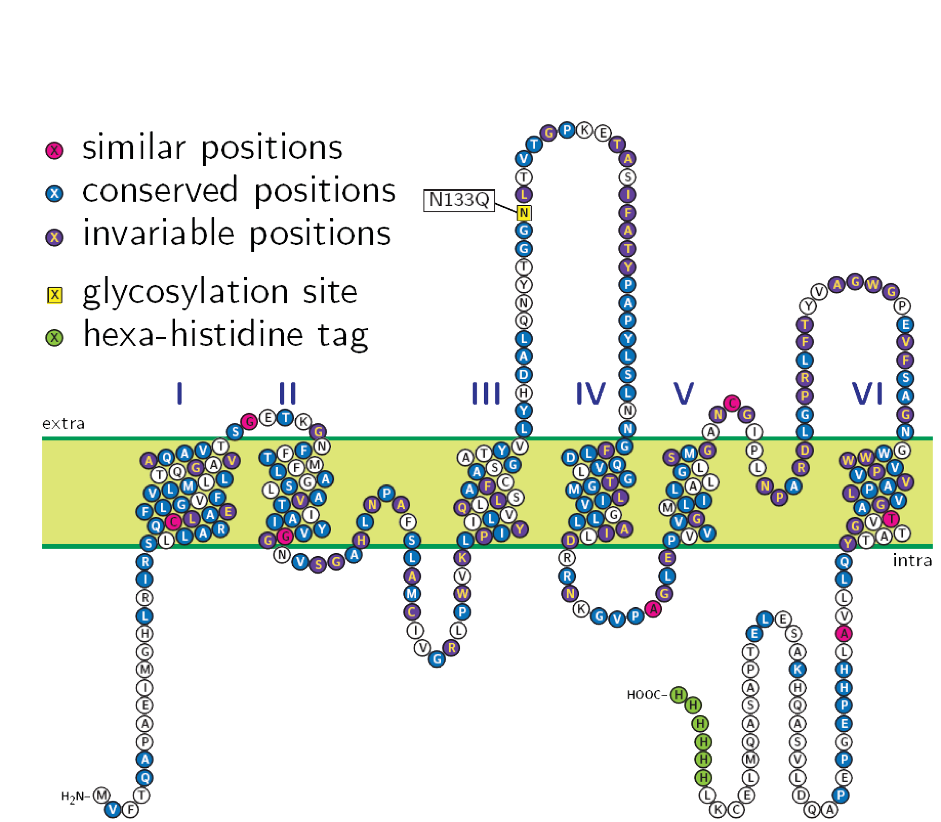
| + | [[Image:100318 All Figures-1.png]] | ||
Current revision
structure of the Plasmodium falciparum aquaporin [1]
Structure of the Escherichia coli aquaglyceroporin [2]
You can clearly see the beta-octyl-glucoside that is bound to the monomer.
|
This is my first attempt.

