Image:1z10pt1.pngj
From Proteopedia
(Difference between revisions)
Size of this preview: 661 × 599 pixels
Full resolution (663 × 601 pixel, file size: 437 KB, MIME type: image/png)
(uploaded a new version of "Image:1z10pt1.pngj") |
(uploaded a new version of "Image:1z10pt1.pngj") |
||
| (One intermediate revision not shown.) | |||
Current revision
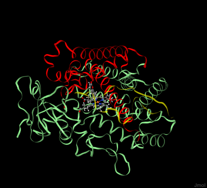
The hCYP 2A6 with highlighted ribbons representing an outline of the active site cavity.
File history
Click on a date/time to view the file as it appeared at that time.
| Date/Time | User | Dimensions | File size | Comment | |
|---|---|---|---|---|---|
| (current) | 20:20, 18 June 2012 | Kellan Passow (Talk | contribs) | 663×601 | 437 KB | |
| 19:17, 18 June 2012 | Kellan Passow (Talk | contribs) | 663×601 | 436 KB | ||
| 15:42, 14 June 2012 | Kellan Passow (Talk | contribs) | 800×596 | 453 KB | ||
| 15:34, 14 June 2012 | Kellan Passow (Talk | contribs) | 800×596 | 464 KB | ||
| 15:19, 14 June 2012 | Kellan Passow (Talk | contribs) | 800×596 | 143 KB | ||
| 20:51, 13 June 2012 | Kellan Passow (Talk | contribs) | 800×598 | 119 KB | The hCYP 2A6 with highlighted ribbons representing an outline of the active site cavity. |
- Edit this file using an external application
See the setup instructions for more information.
Links
There are no pages that link to this file.
