We apologize for Proteopedia being slow to respond. For the past two years, a new implementation of Proteopedia has been being built. Soon, it will replace this 18-year old system. All existing content will be moved to the new system at a date that will be announced here.
3ci0
From Proteopedia
(Difference between revisions)
| (8 intermediate revisions not shown.) | |||
| Line 1: | Line 1: | ||
| - | [[Image:3ci0.png|left|200px]] | ||
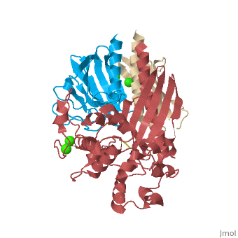
| - | + | ==The Crystal Structure of the GspK-GspI-GspJ complex from enterotoxigenic Escherichia coli Type 2 Secretion System== | |
| + | <StructureSection load='3ci0' size='340' side='right'caption='[[3ci0]], [[Resolution|resolution]] 2.20Å' scene=''> | ||
| + | == Structural highlights == | ||
| + | <table><tr><td colspan='2'>[[3ci0]] is a 3 chain structure with sequence from [https://en.wikipedia.org/wiki/Escherichia_coli Escherichia coli]. Full crystallographic information is available from [http://oca.weizmann.ac.il/oca-bin/ocashort?id=3CI0 OCA]. For a <b>guided tour on the structure components</b> use [https://proteopedia.org/fgij/fg.htm?mol=3CI0 FirstGlance]. <br> | ||
| + | </td></tr><tr id='method'><td class="sblockLbl"><b>[[Empirical_models|Method:]]</b></td><td class="sblockDat" id="methodDat">X-ray diffraction, [[Resolution|Resolution]] 2.2Å</td></tr> | ||
| + | <tr id='ligand'><td class="sblockLbl"><b>[[Ligand|Ligands:]]</b></td><td class="sblockDat" id="ligandDat"><scene name='pdbligand=CA:CALCIUM+ION'>CA</scene>, <scene name='pdbligand=CL:CHLORIDE+ION'>CL</scene>, <scene name='pdbligand=MSE:SELENOMETHIONINE'>MSE</scene></td></tr> | ||
| + | <tr id='resources'><td class="sblockLbl"><b>Resources:</b></td><td class="sblockDat"><span class='plainlinks'>[https://proteopedia.org/fgij/fg.htm?mol=3ci0 FirstGlance], [http://oca.weizmann.ac.il/oca-bin/ocaids?id=3ci0 OCA], [https://pdbe.org/3ci0 PDBe], [https://www.rcsb.org/pdb/explore.do?structureId=3ci0 RCSB], [https://www.ebi.ac.uk/pdbsum/3ci0 PDBsum], [https://prosat.h-its.org/prosat/prosatexe?pdbcode=3ci0 ProSAT]</span></td></tr> | ||
| + | </table> | ||
| + | == Function == | ||
| + | [https://www.uniprot.org/uniprot/GSPI_ECOLX GSPI_ECOLX] Component of the type II secretion system required for the energy-dependent secretion of extracellular factors such as proteases and toxins from the periplasm. Part of the pseudopilus tip complex that is critical for the recognition and binding of secretion substrates.[UniProtKB:Q00516] | ||
| + | == Evolutionary Conservation == | ||
| + | [[Image:Consurf_key_small.gif|200px|right]] | ||
| + | Check<jmol> | ||
| + | <jmolCheckbox> | ||
| + | <scriptWhenChecked>; select protein; define ~consurf_to_do selected; consurf_initial_scene = true; script "/wiki/ConSurf/ci/3ci0_consurf.spt"</scriptWhenChecked> | ||
| + | <scriptWhenUnchecked>script /wiki/extensions/Proteopedia/spt/initialview03.spt</scriptWhenUnchecked> | ||
| + | <text>to colour the structure by Evolutionary Conservation</text> | ||
| + | </jmolCheckbox> | ||
| + | </jmol>, as determined by [http://consurfdb.tau.ac.il/ ConSurfDB]. You may read the [[Conservation%2C_Evolutionary|explanation]] of the method and the full data available from [http://bental.tau.ac.il/new_ConSurfDB/main_output.php?pdb_ID=3ci0 ConSurf]. | ||
| + | <div style="clear:both"></div> | ||
| + | <div style="background-color:#fffaf0;"> | ||
| + | == Publication Abstract from PubMed == | ||
| + | Gram-negative bacteria translocate various proteins including virulence factors across their outer membrane via type 2 secretion systems (T2SSs). T2SSs are thought to contain a pseudopilus, a subcomplex formed by one major and several minor pseudopilins. We report the crystal structure of the complex formed by three minor pseudopilins from enterotoxigenic Escherichia coli. The GspK-GspI-GspJ complex has quasihelical characteristics and an architecture consistent with a localization at the pseudopilus tip. The alpha-domain of GspK has a previously unobserved fold with an unexpected dinuclear metal binding site. The area surrounding its disulfide bridge is conserved and might interact with other T2SS components or with secreted proteins. | ||
| - | + | Structure of the GspK-GspI-GspJ complex from the enterotoxigenic Escherichia coli type 2 secretion system.,Korotkov KV, Hol WG Nat Struct Mol Biol. 2008 May;15(5):462-8. Epub 2008 Apr 27. PMID:18438417<ref>PMID:18438417</ref> | |
| - | + | From MEDLINE®/PubMed®, a database of the U.S. National Library of Medicine.<br> | |
| - | + | </div> | |
| - | + | <div class="pdbe-citations 3ci0" style="background-color:#fffaf0;"></div> | |
| - | + | ||
==See Also== | ==See Also== | ||
*[[Pseudopilin|Pseudopilin]] | *[[Pseudopilin|Pseudopilin]] | ||
| - | + | == References == | |
| - | == | + | <references/> |
| - | < | + | __TOC__ |
| + | </StructureSection> | ||
[[Category: Escherichia coli]] | [[Category: Escherichia coli]] | ||
| - | [[Category: | + | [[Category: Large Structures]] |
| - | [[Category: | + | [[Category: Hol WGJ]] |
| - | [[Category: | + | [[Category: Korotkov KV]] |
| - | + | ||
| - | + | ||
| - | + | ||
| - | + | ||
Current revision
The Crystal Structure of the GspK-GspI-GspJ complex from enterotoxigenic Escherichia coli Type 2 Secretion System
| |||||||||||