This old version of Proteopedia is provided for student assignments while the new version is undergoing repairs. Content and edits done in this old version of Proteopedia after March 1, 2026 will eventually be lost when it is retired in about June of 2026.
Apply for new accounts at the new Proteopedia. Your logins will work in both the old and new versions.
1bm0
From Proteopedia
(Difference between revisions)
| (9 intermediate revisions not shown.) | |||
| Line 1: | Line 1: | ||
| - | [[Image:1bm0.png|left|200px]] | ||
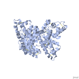
| - | + | ==CRYSTAL STRUCTURE OF HUMAN SERUM ALBUMIN== | |
| + | <StructureSection load='1bm0' size='340' side='right'caption='[[1bm0]], [[Resolution|resolution]] 2.50Å' scene=''> | ||
| + | == Structural highlights == | ||
| + | <table><tr><td colspan='2'>[[1bm0]] is a 2 chain structure with sequence from [https://en.wikipedia.org/wiki/Homo_sapiens Homo sapiens]. Full crystallographic information is available from [http://oca.weizmann.ac.il/oca-bin/ocashort?id=1BM0 OCA]. For a <b>guided tour on the structure components</b> use [https://proteopedia.org/fgij/fg.htm?mol=1BM0 FirstGlance]. <br> | ||
| + | </td></tr><tr id='method'><td class="sblockLbl"><b>[[Empirical_models|Method:]]</b></td><td class="sblockDat" id="methodDat">X-ray diffraction, [[Resolution|Resolution]] 2.5Å</td></tr> | ||
| + | <tr id='resources'><td class="sblockLbl"><b>Resources:</b></td><td class="sblockDat"><span class='plainlinks'>[https://proteopedia.org/fgij/fg.htm?mol=1bm0 FirstGlance], [http://oca.weizmann.ac.il/oca-bin/ocaids?id=1bm0 OCA], [https://pdbe.org/1bm0 PDBe], [https://www.rcsb.org/pdb/explore.do?structureId=1bm0 RCSB], [https://www.ebi.ac.uk/pdbsum/1bm0 PDBsum], [https://prosat.h-its.org/prosat/prosatexe?pdbcode=1bm0 ProSAT]</span></td></tr> | ||
| + | </table> | ||
| + | == Disease == | ||
| + | [https://www.uniprot.org/uniprot/ALBU_HUMAN ALBU_HUMAN] Defects in ALB are a cause of familial dysalbuminemic hyperthyroxinemia (FDH) [MIM:[https://omim.org/entry/103600 103600]. FDH is a form of euthyroid hyperthyroxinemia that is due to increased affinity of ALB for T(4). It is the most common cause of inherited euthyroid hyperthyroxinemia in Caucasian population.<ref>PMID:8048949</ref> <ref>PMID:7852505</ref> <ref>PMID:9329347</ref> <ref>PMID:9589637</ref> | ||
| + | == Function == | ||
| + | [https://www.uniprot.org/uniprot/ALBU_HUMAN ALBU_HUMAN] Serum albumin, the main protein of plasma, has a good binding capacity for water, Ca(2+), Na(+), K(+), fatty acids, hormones, bilirubin and drugs. Its main function is the regulation of the colloidal osmotic pressure of blood. Major zinc transporter in plasma, typically binds about 80% of all plasma zinc.<ref>PMID:19021548</ref> | ||
| + | == Evolutionary Conservation == | ||
| + | [[Image:Consurf_key_small.gif|200px|right]] | ||
| + | Check<jmol> | ||
| + | <jmolCheckbox> | ||
| + | <scriptWhenChecked>; select protein; define ~consurf_to_do selected; consurf_initial_scene = true; script "/wiki/ConSurf/bm/1bm0_consurf.spt"</scriptWhenChecked> | ||
| + | <scriptWhenUnchecked>script /wiki/extensions/Proteopedia/spt/initialview01.spt</scriptWhenUnchecked> | ||
| + | <text>to colour the structure by Evolutionary Conservation</text> | ||
| + | </jmolCheckbox> | ||
| + | </jmol>, as determined by [http://consurfdb.tau.ac.il/ ConSurfDB]. You may read the [[Conservation%2C_Evolutionary|explanation]] of the method and the full data available from [http://bental.tau.ac.il/new_ConSurfDB/main_output.php?pdb_ID=1bm0 ConSurf]. | ||
| + | <div style="clear:both"></div> | ||
| + | <div style="background-color:#fffaf0;"> | ||
| + | == Publication Abstract from PubMed == | ||
| + | A new triclinic crystal form of human serum albumin (HSA), derived either from pool plasma (pHSA) or from a Pichia pastoris expression system (rHSA), was obtained from polyethylene glycol 4000 solution. Three-dimensional structures of pHSA and rHSA were determined at 2.5 A resolution from the new triclinic crystal form by molecular replacement, using atomic coordinates derived from a multiple isomorphous replacement work with a known tetragonal crystal form. The structures of pHSA and rHSA are virtually identical, with an r.m. s. deviation of 0.24 A for all Calpha atoms. The two HSA molecules involved in the asymmetric unit are related by a strict local twofold symmetry such that the Calpha atoms of the two molecules can be superimposed with an r.m.s. deviation of 0.28 A in pHSA. Cys34 is the only cysteine with a free sulfhydryl group which does not participate in a disulfide linkage with any external ligand. Domains II and III both have a pocket formed mostly of hydrophobic and positively charged residues and in which a very wide range of compounds may be accommodated. Three tentative binding sites for long-chain fatty acids, each with different surroundings, are located at the surface of each domain. | ||
| - | + | Crystal structure of human serum albumin at 2.5 A resolution.,Sugio S, Kashima A, Mochizuki S, Noda M, Kobayashi K Protein Eng. 1999 Jun;12(6):439-46. PMID:10388840<ref>PMID:10388840</ref> | |
| - | + | From MEDLINE®/PubMed®, a database of the U.S. National Library of Medicine.<br> | |
| - | + | </div> | |
| - | + | <div class="pdbe-citations 1bm0" style="background-color:#fffaf0;"></div> | |
| - | + | ||
==See Also== | ==See Also== | ||
*[[Albumin|Albumin]] | *[[Albumin|Albumin]] | ||
| - | + | *[[Albumin 3D structures|Albumin 3D structures]] | |
| - | == | + | == References == |
| - | < | + | <references/> |
| + | __TOC__ | ||
| + | </StructureSection> | ||
[[Category: Homo sapiens]] | [[Category: Homo sapiens]] | ||
| - | [[Category: Kashima | + | [[Category: Large Structures]] |
| - | [[Category: Kobayashi | + | [[Category: Kashima A]] |
| - | [[Category: Mochizuki | + | [[Category: Kobayashi K]] |
| - | [[Category: Noda | + | [[Category: Mochizuki S]] |
| - | [[Category: Sugio | + | [[Category: Noda M]] |
| - | + | [[Category: Sugio S]] | |
| - | + | ||
Current revision
CRYSTAL STRUCTURE OF HUMAN SERUM ALBUMIN
| |||||||||||