This old version of Proteopedia is provided for student assignments while the new version is undergoing repairs. Content and edits done in this old version of Proteopedia after March 1, 2026 will eventually be lost when it is retired in about June of 2026.
Apply for new accounts at the new Proteopedia. Your logins will work in both the old and new versions.
2ah4
From Proteopedia
(Difference between revisions)
m (Protected "2ah4" [edit=sysop:move=sysop]) |
|||
| (7 intermediate revisions not shown.) | |||
| Line 1: | Line 1: | ||
| - | [[Image:2ah4.png|left|200px]] | ||
| - | + | ==guanidinobenzoyl-trypsin acyl-enzyme at 1.13 A resolution== | |
| + | <StructureSection load='2ah4' size='340' side='right'caption='[[2ah4]], [[Resolution|resolution]] 1.13Å' scene=''> | ||
| + | == Structural highlights == | ||
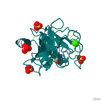
| + | <table><tr><td colspan='2'>[[2ah4]] is a 1 chain structure with sequence from [https://en.wikipedia.org/wiki/Bos_taurus Bos taurus]. Full crystallographic information is available from [http://oca.weizmann.ac.il/oca-bin/ocashort?id=2AH4 OCA]. For a <b>guided tour on the structure components</b> use [https://proteopedia.org/fgij/fg.htm?mol=2AH4 FirstGlance]. <br> | ||
| + | </td></tr><tr id='method'><td class="sblockLbl"><b>[[Empirical_models|Method:]]</b></td><td class="sblockDat" id="methodDat">X-ray diffraction, [[Resolution|Resolution]] 1.13Å</td></tr> | ||
| + | <tr id='ligand'><td class="sblockLbl"><b>[[Ligand|Ligands:]]</b></td><td class="sblockDat" id="ligandDat"><scene name='pdbligand=CA:CALCIUM+ION'>CA</scene>, <scene name='pdbligand=GBS:4-GUANIDINOBENZOIC+ACID'>GBS</scene>, <scene name='pdbligand=SO4:SULFATE+ION'>SO4</scene></td></tr> | ||
| + | <tr id='resources'><td class="sblockLbl"><b>Resources:</b></td><td class="sblockDat"><span class='plainlinks'>[https://proteopedia.org/fgij/fg.htm?mol=2ah4 FirstGlance], [http://oca.weizmann.ac.il/oca-bin/ocaids?id=2ah4 OCA], [https://pdbe.org/2ah4 PDBe], [https://www.rcsb.org/pdb/explore.do?structureId=2ah4 RCSB], [https://www.ebi.ac.uk/pdbsum/2ah4 PDBsum], [https://prosat.h-its.org/prosat/prosatexe?pdbcode=2ah4 ProSAT]</span></td></tr> | ||
| + | </table> | ||
| + | == Function == | ||
| + | [https://www.uniprot.org/uniprot/TRY1_BOVIN TRY1_BOVIN] | ||
| + | == Evolutionary Conservation == | ||
| + | [[Image:Consurf_key_small.gif|200px|right]] | ||
| + | Check<jmol> | ||
| + | <jmolCheckbox> | ||
| + | <scriptWhenChecked>; select protein; define ~consurf_to_do selected; consurf_initial_scene = true; script "/wiki/ConSurf/ah/2ah4_consurf.spt"</scriptWhenChecked> | ||
| + | <scriptWhenUnchecked>script /wiki/extensions/Proteopedia/spt/initialview01.spt</scriptWhenUnchecked> | ||
| + | <text>to colour the structure by Evolutionary Conservation</text> | ||
| + | </jmolCheckbox> | ||
| + | </jmol>, as determined by [http://consurfdb.tau.ac.il/ ConSurfDB]. You may read the [[Conservation%2C_Evolutionary|explanation]] of the method and the full data available from [http://bental.tau.ac.il/new_ConSurfDB/main_output.php?pdb_ID=2ah4 ConSurf]. | ||
| + | <div style="clear:both"></div> | ||
| + | <div style="background-color:#fffaf0;"> | ||
| + | == Publication Abstract from PubMed == | ||
| + | Atomic resolution structures of trypsin acyl-enzymes and a tetrahedral intermediate analog, along with previously solved structures representing the Michaelis complex, are used to reconstruct events in the catalytic cycle of this classic serine protease. Structural comparisons provide insight into active site adjustments involved in catalysis. Subtle motions of the catalytic serine and histidine residues coordinated with translation of the substrate reaction center are seen to favor the forward progress of the acylation reaction. The structures also clarify the attack trajectory of the hydrolytic water in the deacylation reaction. | ||
| - | + | Insights into the serine protease mechanism from atomic resolution structures of trypsin reaction intermediates.,Radisky ES, Lee JM, Lu CJ, Koshland DE Jr Proc Natl Acad Sci U S A. 2006 May 2;103(18):6835-40. Epub 2006 Apr 24. PMID:16636277<ref>PMID:16636277</ref> | |
| - | + | From MEDLINE®/PubMed®, a database of the U.S. National Library of Medicine.<br> | |
| - | + | </div> | |
| - | + | <div class="pdbe-citations 2ah4" style="background-color:#fffaf0;"></div> | |
| - | + | ||
==See Also== | ==See Also== | ||
| - | *[[Trypsin|Trypsin]] | + | *[[Trypsin 3D structures|Trypsin 3D structures]] |
| - | + | == References == | |
| - | == | + | <references/> |
| - | < | + | __TOC__ |
| + | </StructureSection> | ||
[[Category: Bos taurus]] | [[Category: Bos taurus]] | ||
| - | [[Category: | + | [[Category: Large Structures]] |
| - | [[Category: Koshland | + | [[Category: Koshland Jr DE]] |
| - | [[Category: Lee | + | [[Category: Lee JM]] |
| - | [[Category: Lu | + | [[Category: Lu CJ]] |
| - | [[Category: Radisky | + | [[Category: Radisky ES]] |
| - | + | ||
| - | + | ||
| - | + | ||
| - | + | ||
| - | + | ||
Current revision
guanidinobenzoyl-trypsin acyl-enzyme at 1.13 A resolution
| |||||||||||