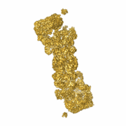
Sandbox pia
From Proteopedia
| (5 intermediate revisions not shown.) | |||
| Line 1: | Line 1: | ||
== Structural model of yeast 26S == | == Structural model of yeast 26S == | ||
| - | |||
| - | |||
==About this Structure== | ==About this Structure== | ||
[[4b4t]] is a 31 chain structure with sequence from [http://en.wikipedia.org/wiki/Saccharomyces_cerevisiae Saccharomyces cerevisiae]. Full crystallographic information is available from [http://oca.weizmann.ac.il/oca-bin/ocashort?id=4B4T OCA] and described in Beck ''et al.''<ref name="Beck">pmid 22927375</ref>. The structure is a comparative model which was fitted into an cryo-EM density at 7.4Å resolution. Previous publications describing the architecture are <ref>pmid 22307589</ref> <ref>pmid 22237024 </ref> <ref>pmid 20467039 </ref> | [[4b4t]] is a 31 chain structure with sequence from [http://en.wikipedia.org/wiki/Saccharomyces_cerevisiae Saccharomyces cerevisiae]. Full crystallographic information is available from [http://oca.weizmann.ac.il/oca-bin/ocashort?id=4B4T OCA] and described in Beck ''et al.''<ref name="Beck">pmid 22927375</ref>. The structure is a comparative model which was fitted into an cryo-EM density at 7.4Å resolution. Previous publications describing the architecture are <ref>pmid 22307589</ref> <ref>pmid 22237024 </ref> <ref>pmid 20467039 </ref> | ||
| - | <StructureSection load='4b4t' size='350' side='right' caption='Structural model of yeast 26S (PDB entry [[4b4t]])' scene='Sandbox_pia/26s_color/ | + | <StructureSection load='4b4t' size='350' side='right' caption='Structural model of yeast 26S (PDB entry [[4b4t]])' scene='Sandbox_pia/26s_color/3'> |
The model displays one half of the symmetric 26S proteasome <scene name='Sandbox_pia/26s_color/1'>26S</scene>. The complex consists of a core particle, build out of 4 heptameric rings in the order α - β - β - α (<scene name='Sandbox_pia/Cp/1'>half CP</scene>)<br> | The model displays one half of the symmetric 26S proteasome <scene name='Sandbox_pia/26s_color/1'>26S</scene>. The complex consists of a core particle, build out of 4 heptameric rings in the order α - β - β - α (<scene name='Sandbox_pia/Cp/1'>half CP</scene>)<br> | ||
| - | The regulatory particle is divided into a base and a lid. The base are 6 AAA-ATPases (Rpt1-6) | + | The regulatory particle is divided into a base and a lid. The base are 6 <scene name='Sandbox_pia/Aaa/3'>AAA-ATPases </scene> (Rpt1-6) and Rpn1 whereas the lid consists of non-ATPases, some containing a <scene name='Sandbox_pia/Pci/2'>PCI </scene> domain( Rpn9,5,6,7,3,12) or a <scene name='Sandbox_pia/Mpn/5'>MPN</scene> domain (Rpn8, 11) as well as Ubiquitin receptors (Rpn10,13).<br> |
| - | The C-termini (like the <scene name='Sandbox_pia/Rpt2_ter/3'>Rpt2 C-Terminus</scene>) of some ATPases can be localized in pockets between the alpha subunits and might play a role in gate opening, as suggested by <ref> pmid </ref>. | + | The C-termini (like the <scene name='Sandbox_pia/Rpt2_ter/3'>Rpt2 C-Terminus</scene>) of some ATPases can be localized in pockets between the alpha subunits and might play a role in gate opening, as suggested by <ref> pmid 17803938</ref>. |
| - | + | Moreover, the <scene name='Sandbox_pia/Walker_motifs/1'>Walker motifs</scene> of the AAA-ATPases, modeled based on PAN [[3h4m]] are conserved. | |
</StructureSection> | </StructureSection> | ||
| Line 18: | Line 16: | ||
The 26S proteasome operates at the executive end of the ubiquitin-proteasome | The 26S proteasome operates at the executive end of the ubiquitin-proteasome | ||
pathway. Here, we present a cryo-EM structure of the | pathway. Here, we present a cryo-EM structure of the | ||
| - | Saccharomyces cerevisiae 26S proteasome at a resolution of 7.4 Å or 6.7 Å (Fourier-Shell Correlation of 0.5 or 0.3, respectively). [[Image:26S.gif|thumb|Cryo-EM density [ | + | Saccharomyces cerevisiae 26S proteasome at a resolution of 7.4 Å or 6.7 Å (Fourier-Shell Correlation of 0.5 or 0.3, respectively). [[Image:26S.gif|thumb|Cryo-EM density [http://www.ebi.ac.uk/pdbe-srv/emsearch/atlas/2165_visualization.html emdb-2165] used for flexible fitting with MDFF<ref>pmid 19398010</ref> <ref name="Beck"/> ]] |
We used this map in conjunction with molecular dynamics-based flexible | We used this map in conjunction with molecular dynamics-based flexible | ||
fitting to build a near-atomic resolution model of the holocomplex. | fitting to build a near-atomic resolution model of the holocomplex. | ||
| Line 39: | Line 37: | ||
| - | == References == | + | ==References== |
<references/> | <references/> | ||
Current revision
Structural model of yeast 26S
About this Structure
4b4t is a 31 chain structure with sequence from Saccharomyces cerevisiae. Full crystallographic information is available from OCA and described in Beck et al.[1]. The structure is a comparative model which was fitted into an cryo-EM density at 7.4Å resolution. Previous publications describing the architecture are [2] [3] [4]
| |||||||||||
Abstract Beck et al.[1]:
The 26S proteasome operates at the executive end of the ubiquitin-proteasome
pathway. Here, we present a cryo-EM structure of the
We used this map in conjunction with molecular dynamics-based flexible fitting to build a near-atomic resolution model of the holocomplex. The quality of the map allowed us to assign α-helices, the predominant secondary structure element of the regulatory particle subunits, throughout the entire map. We were able to determine the architecture of the Rpn8/Rpn11 heterodimer, which had hitherto remained elusive. The MPN domain of Rpn11 is positioned directly above the AAA-ATPase N-ring suggesting that Rpn11 deubiquitylates substrates immediately following commitment and prior to their unfolding by the AAA-ATPase module. The MPN domain of Rpn11 dimerizes with that of Rpn8 and the C-termini of both subunits form long helices, which are integral parts of a coiled-coil module. Together with the C-terminal helices of the six PCI-domain subunits they form a very large coiled-coil bundle, which appears to serve as a flexible anchoring device for all the lid subunits.
References
- ↑ 1.0 1.1 1.2 Beck F, Unverdorben P, Bohn S, Schweitzer A, Pfeifer G, Sakata E, Nickell S, Plitzko JM, Villa E, Baumeister W, Forster F. Near-atomic resolution structural model of the yeast 26S proteasome. Proc Natl Acad Sci U S A. 2012 Aug 27. PMID:22927375 doi:10.1073/pnas.1213333109
- ↑ Lasker K, Forster F, Bohn S, Walzthoeni T, Villa E, Unverdorben P, Beck F, Aebersold R, Sali A, Baumeister W. Molecular architecture of the 26S proteasome holocomplex determined by an integrative approach. Proc Natl Acad Sci U S A. 2012 Jan 31;109(5):1380-7. Epub 2012 Jan 23. PMID:22307589 doi:10.1073/pnas.1120559109
- ↑ Lander GC, Estrin E, Matyskiela ME, Bashore C, Nogales E, Martin A. Complete subunit architecture of the proteasome regulatory particle. Nature. 2012 Jan 11;482(7384):186-91. doi: 10.1038/nature10774. PMID:22237024 doi:10.1038/nature10774
- ↑ Forster F, Lasker K, Nickell S, Sali A, Baumeister W. Toward an integrated structural model of the 26S proteasome. Mol Cell Proteomics. 2010 Aug;9(8):1666-77. Epub 2010 May 13. PMID:20467039 doi:10.1074/mcp.R000002-MCP201
- ↑ Smith DM, Chang SC, Park S, Finley D, Cheng Y, Goldberg AL. Docking of the proteasomal ATPases' carboxyl termini in the 20S proteasome's alpha ring opens the gate for substrate entry. Mol Cell. 2007 Sep 7;27(5):731-44. PMID:17803938 doi:10.1016/j.molcel.2007.06.033
- ↑ Trabuco LG, Villa E, Schreiner E, Harrison CB, Schulten K. Molecular dynamics flexible fitting: a practical guide to combine cryo-electron microscopy and X-ray crystallography. Methods. 2009 Oct;49(2):174-80. Epub 2009 May 4. PMID:19398010 doi:10.1016/j.ymeth.2009.04.005

