This old version of Proteopedia is provided for student assignments while the new version is undergoing repairs. Content and edits done in this old version of Proteopedia after March 1, 2026 will eventually be lost when it is retired in about June of 2026.
Apply for new accounts at the new Proteopedia. Your logins will work in both the old and new versions.
1prt
From Proteopedia
(Difference between revisions)
| (7 intermediate revisions not shown.) | |||
| Line 1: | Line 1: | ||
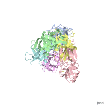
| - | [[Image:1prt.png|left|200px]] | ||
| - | + | ==THE CRYSTAL STRUCTURE OF PERTUSSIS TOXIN== | |
| - | + | <StructureSection load='1prt' size='340' side='right'caption='[[1prt]], [[Resolution|resolution]] 2.90Å' scene=''> | |
| - | + | == Structural highlights == | |
| - | + | <table><tr><td colspan='2'>[[1prt]] is a 12 chain structure with sequence from [https://en.wikipedia.org/wiki/Bordetella_pertussis Bordetella pertussis]. The September 2005 RCSB PDB [https://pdb.rcsb.org/pdb/static.do?p=education_discussion/molecule_of_the_month/index.html Molecule of the Month] feature on ''Cholera Toxin'' by David S. Goodsell is [https://dx.doi.org/10.2210/rcsb_pdb/mom_2005_9 10.2210/rcsb_pdb/mom_2005_9]. Full crystallographic information is available from [http://oca.weizmann.ac.il/oca-bin/ocashort?id=1PRT OCA]. For a <b>guided tour on the structure components</b> use [https://proteopedia.org/fgij/fg.htm?mol=1PRT FirstGlance]. <br> | |
| - | + | </td></tr><tr id='method'><td class="sblockLbl"><b>[[Empirical_models|Method:]]</b></td><td class="sblockDat" id="methodDat">X-ray diffraction, [[Resolution|Resolution]] 2.9Å</td></tr> | |
| - | + | <tr id='resources'><td class="sblockLbl"><b>Resources:</b></td><td class="sblockDat"><span class='plainlinks'>[https://proteopedia.org/fgij/fg.htm?mol=1prt FirstGlance], [http://oca.weizmann.ac.il/oca-bin/ocaids?id=1prt OCA], [https://pdbe.org/1prt PDBe], [https://www.rcsb.org/pdb/explore.do?structureId=1prt RCSB], [https://www.ebi.ac.uk/pdbsum/1prt PDBsum], [https://prosat.h-its.org/prosat/prosatexe?pdbcode=1prt ProSAT]</span></td></tr> | |
| - | == | + | </table> |
| - | [[1prt]] is a 12 chain structure with sequence from [ | + | == Function == |
| + | [https://www.uniprot.org/uniprot/TOX1_BORPE TOX1_BORPE] S1 is an NAD-dependent ADP-ribosyltransferase, which plays a crucial role in the pathogenesis of B.pertussis causing disruption of normal host cellular regulation. It catalyzes the ADP-ribosylation of a cysteine in the alpha subunit of host heterotrimeric G proteins. In the absence of G proteins it also catalyzes the cleavage of NAD(+) into ADP-ribose and nicotinamide. It irreversibly uncouples the G-alpha GTP-binding proteins from their membrane receptors. | ||
| + | == Evolutionary Conservation == | ||
| + | [[Image:Consurf_key_small.gif|200px|right]] | ||
| + | Check<jmol> | ||
| + | <jmolCheckbox> | ||
| + | <scriptWhenChecked>; select protein; define ~consurf_to_do selected; consurf_initial_scene = true; script "/wiki/ConSurf/pr/1prt_consurf.spt"</scriptWhenChecked> | ||
| + | <scriptWhenUnchecked>script /wiki/extensions/Proteopedia/spt/initialview01.spt</scriptWhenUnchecked> | ||
| + | <text>to colour the structure by Evolutionary Conservation</text> | ||
| + | </jmolCheckbox> | ||
| + | </jmol>, as determined by [http://consurfdb.tau.ac.il/ ConSurfDB]. You may read the [[Conservation%2C_Evolutionary|explanation]] of the method and the full data available from [http://bental.tau.ac.il/new_ConSurfDB/main_output.php?pdb_ID=1prt ConSurf]. | ||
| + | <div style="clear:both"></div> | ||
==See Also== | ==See Also== | ||
*[[Pertussis toxin|Pertussis toxin]] | *[[Pertussis toxin|Pertussis toxin]] | ||
| - | + | __TOC__ | |
| - | + | </StructureSection> | |
| - | + | ||
[[Category: Bordetella pertussis]] | [[Category: Bordetella pertussis]] | ||
[[Category: Cholera Toxin]] | [[Category: Cholera Toxin]] | ||
| + | [[Category: Large Structures]] | ||
[[Category: RCSB PDB Molecule of the Month]] | [[Category: RCSB PDB Molecule of the Month]] | ||
| - | [[Category: Read | + | [[Category: Read RJ]] |
| - | [[Category: Stein | + | [[Category: Stein PE]] |
| - | + | ||
Current revision
THE CRYSTAL STRUCTURE OF PERTUSSIS TOXIN
| |||||||||||