This old version of Proteopedia is provided for student assignments while the new version is undergoing repairs. Content and edits done in this old version of Proteopedia after March 1, 2026 will eventually be lost when it is retired in about June of 2026.
Apply for new accounts at the new Proteopedia. Your logins will work in both the old and new versions.
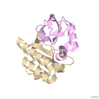
2dfx
From Proteopedia
(Difference between revisions)
| (7 intermediate revisions not shown.) | |||
| Line 1: | Line 1: | ||
| - | [[Image:2dfx.png|left|200px]] | ||
| - | + | ==Crystal structure of the carboxy terminal domain of colicin E5 complexed with its inhibitor== | |
| - | + | <StructureSection load='2dfx' size='340' side='right'caption='[[2dfx]], [[Resolution|resolution]] 1.90Å' scene=''> | |
| - | + | == Structural highlights == | |
| - | + | <table><tr><td colspan='2'>[[2dfx]] is a 2 chain structure with sequence from [https://en.wikipedia.org/wiki/Escherichia_coli Escherichia coli]. Full crystallographic information is available from [http://oca.weizmann.ac.il/oca-bin/ocashort?id=2DFX OCA]. For a <b>guided tour on the structure components</b> use [https://proteopedia.org/fgij/fg.htm?mol=2DFX FirstGlance]. <br> | |
| - | + | </td></tr><tr id='method'><td class="sblockLbl"><b>[[Empirical_models|Method:]]</b></td><td class="sblockDat" id="methodDat">X-ray diffraction, [[Resolution|Resolution]] 1.9Å</td></tr> | |
| - | + | <tr id='resources'><td class="sblockLbl"><b>Resources:</b></td><td class="sblockDat"><span class='plainlinks'>[https://proteopedia.org/fgij/fg.htm?mol=2dfx FirstGlance], [http://oca.weizmann.ac.il/oca-bin/ocaids?id=2dfx OCA], [https://pdbe.org/2dfx PDBe], [https://www.rcsb.org/pdb/explore.do?structureId=2dfx RCSB], [https://www.ebi.ac.uk/pdbsum/2dfx PDBsum], [https://prosat.h-its.org/prosat/prosatexe?pdbcode=2dfx ProSAT]</span></td></tr> | |
| - | == | + | </table> |
| - | [[2dfx]] is a 2 chain structure with sequence from [ | + | == Function == |
| + | [https://www.uniprot.org/uniprot/CEA5_ECOLX CEA5_ECOLX] Colicins are polypeptide toxins produced by and active against E.coli and closely related bacteria. This colicin is an endonuclease. | ||
| + | == Evolutionary Conservation == | ||
| + | [[Image:Consurf_key_small.gif|200px|right]] | ||
| + | Check<jmol> | ||
| + | <jmolCheckbox> | ||
| + | <scriptWhenChecked>; select protein; define ~consurf_to_do selected; consurf_initial_scene = true; script "/wiki/ConSurf/df/2dfx_consurf.spt"</scriptWhenChecked> | ||
| + | <scriptWhenUnchecked>script /wiki/extensions/Proteopedia/spt/initialview01.spt</scriptWhenUnchecked> | ||
| + | <text>to colour the structure by Evolutionary Conservation</text> | ||
| + | </jmolCheckbox> | ||
| + | </jmol>, as determined by [http://consurfdb.tau.ac.il/ ConSurfDB]. You may read the [[Conservation%2C_Evolutionary|explanation]] of the method and the full data available from [http://bental.tau.ac.il/new_ConSurfDB/main_output.php?pdb_ID=2dfx ConSurf]. | ||
| + | <div style="clear:both"></div> | ||
==See Also== | ==See Also== | ||
| - | *[[Colicin|Colicin]] | + | *[[Colicin 3D structures|Colicin 3D structures]] |
| - | *[[Colicin | + | *[[Colicin immunity protein 3D structures|Colicin immunity protein 3D structures]] |
| - | + | __TOC__ | |
| - | + | </StructureSection> | |
| - | + | ||
[[Category: Escherichia coli]] | [[Category: Escherichia coli]] | ||
| - | [[Category: Inoue | + | [[Category: Large Structures]] |
| - | [[Category: Masaki | + | [[Category: Inoue S]] |
| - | [[Category: Nonaka | + | [[Category: Masaki H]] |
| - | [[Category: Ogawa | + | [[Category: Nonaka T]] |
| - | [[Category: Ohsawa | + | [[Category: Ogawa T]] |
| - | [[Category: Yajima | + | [[Category: Ohsawa K]] |
| - | + | [[Category: Yajima S]] | |
| - | + | ||
| - | + | ||
Current revision
Crystal structure of the carboxy terminal domain of colicin E5 complexed with its inhibitor
| |||||||||||

Categories: Escherichia coli | Large Structures | Inoue S | Masaki H | Nonaka T | Ogawa T | Ohsawa K | Yajima S
