2jgd
From Proteopedia
(Difference between revisions)
| (6 intermediate revisions not shown.) | |||
| Line 1: | Line 1: | ||
| - | [[Image:2jgd.png|left|200px]] | ||
| - | + | ==E. COLI 2-oxoglutarate dehydrogenase (E1o)== | |
| + | <StructureSection load='2jgd' size='340' side='right'caption='[[2jgd]], [[Resolution|resolution]] 2.60Å' scene=''> | ||
| + | == Structural highlights == | ||
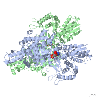
| + | <table><tr><td colspan='2'>[[2jgd]] is a 2 chain structure with sequence from [https://en.wikipedia.org/wiki/Escherichia_coli_K-12 Escherichia coli K-12]. The October 2012 RCSB PDB [https://pdb.rcsb.org/pdb/static.do?p=education_discussion/molecule_of_the_month/index.html Molecule of the Month] feature on ''Citric Acid Cycle'' by David Goodsell is [https://dx.doi.org/10.2210/rcsb_pdb/mom_2012_10 10.2210/rcsb_pdb/mom_2012_10]. Full crystallographic information is available from [http://oca.weizmann.ac.il/oca-bin/ocashort?id=2JGD OCA]. For a <b>guided tour on the structure components</b> use [https://proteopedia.org/fgij/fg.htm?mol=2JGD FirstGlance]. <br> | ||
| + | </td></tr><tr id='method'><td class="sblockLbl"><b>[[Empirical_models|Method:]]</b></td><td class="sblockDat" id="methodDat">X-ray diffraction, [[Resolution|Resolution]] 2.6Å</td></tr> | ||
| + | <tr id='ligand'><td class="sblockLbl"><b>[[Ligand|Ligands:]]</b></td><td class="sblockDat" id="ligandDat"><scene name='pdbligand=AMP:ADENOSINE+MONOPHOSPHATE'>AMP</scene></td></tr> | ||
| + | <tr id='resources'><td class="sblockLbl"><b>Resources:</b></td><td class="sblockDat"><span class='plainlinks'>[https://proteopedia.org/fgij/fg.htm?mol=2jgd FirstGlance], [http://oca.weizmann.ac.il/oca-bin/ocaids?id=2jgd OCA], [https://pdbe.org/2jgd PDBe], [https://www.rcsb.org/pdb/explore.do?structureId=2jgd RCSB], [https://www.ebi.ac.uk/pdbsum/2jgd PDBsum], [https://prosat.h-its.org/prosat/prosatexe?pdbcode=2jgd ProSAT]</span></td></tr> | ||
| + | </table> | ||
| + | == Function == | ||
| + | [https://www.uniprot.org/uniprot/ODO1_ECOLI ODO1_ECOLI] The 2-oxoglutarate dehydrogenase complex catalyzes the overall conversion of 2-oxoglutarate to succinyl-CoA and CO(2). It contains multiple copies of three enzymatic components: 2-oxoglutarate dehydrogenase (E1), dihydrolipoamide succinyltransferase (E2) and lipoamide dehydrogenase (E3). | ||
| + | == Evolutionary Conservation == | ||
| + | [[Image:Consurf_key_small.gif|200px|right]] | ||
| + | Check<jmol> | ||
| + | <jmolCheckbox> | ||
| + | <scriptWhenChecked>; select protein; define ~consurf_to_do selected; consurf_initial_scene = true; script "/wiki/ConSurf/jg/2jgd_consurf.spt"</scriptWhenChecked> | ||
| + | <scriptWhenUnchecked>script /wiki/extensions/Proteopedia/spt/initialview01.spt</scriptWhenUnchecked> | ||
| + | <text>to colour the structure by Evolutionary Conservation</text> | ||
| + | </jmolCheckbox> | ||
| + | </jmol>, as determined by [http://consurfdb.tau.ac.il/ ConSurfDB]. You may read the [[Conservation%2C_Evolutionary|explanation]] of the method and the full data available from [http://bental.tau.ac.il/new_ConSurfDB/main_output.php?pdb_ID=2jgd ConSurf]. | ||
| + | <div style="clear:both"></div> | ||
| + | <div style="background-color:#fffaf0;"> | ||
| + | == Publication Abstract from PubMed == | ||
| + | The thiamine-dependent E1o component (EC 1.2.4.2) of the 2-oxoglutarate dehydrogenase complex catalyses a rate-limiting step of the tricarboxylic acid cycle (TCA) of aerobically respiring organisms. We describe the crystal structure of Escherichia coli E1o in its apo and holo forms at 2.6 A and 3.5 A resolution, respectively. The structures reveal the characteristic fold that binds thiamine diphosphate and resemble closely the alpha(2)beta(2) hetero-tetrameric E1 components of other 2-oxo acid dehydrogenase complexes, except that in E1o, the alpha and beta subunits are fused as a single polypeptide. The extended segment that links the alpha-like and beta-like domains forms a pocket occupied by AMP, which is recognised specifically. Also distinctive to E1o are N-terminal extensions to the core fold, and which may mediate interactions with other components of the 2-oxoglutarate dehydrogenase multienzyme complex. The active site pocket contains a group of three histidine residues and one serine that appear to confer substrate specificity and the capacity to accommodate the TCA metabolite oxaloacetate. Oxaloacetate inhibits E1o activity at physiological concentrations, and we suggest that the inhibition may allow coordinated activity within the TCA cycle. We discuss the implications for metabolic control in facultative anaerobes, and for energy homeostasis of the mammalian brain. | ||
| - | + | Crystal structure of the E1 component of the Escherichia coli 2-oxoglutarate dehydrogenase multienzyme complex.,Frank RA, Price AJ, Northrop FD, Perham RN, Luisi BF J Mol Biol. 2007 May 4;368(3):639-51. Epub 2007 Feb 7. PMID:17367808<ref>PMID:17367808</ref> | |
| - | + | From MEDLINE®/PubMed®, a database of the U.S. National Library of Medicine.<br> | |
| - | + | </div> | |
| - | + | <div class="pdbe-citations 2jgd" style="background-color:#fffaf0;"></div> | |
| - | + | ||
==See Also== | ==See Also== | ||
*[[2-Oxoglutarate Dehydrogenase|2-Oxoglutarate Dehydrogenase]] | *[[2-Oxoglutarate Dehydrogenase|2-Oxoglutarate Dehydrogenase]] | ||
| - | + | *[[2-oxoglutarate dehydrogenase 3D structures|2-oxoglutarate dehydrogenase 3D structures]] | |
| - | == | + | == References == |
| - | < | + | <references/> |
| - | [[Category: | + | __TOC__ |
| - | [[Category: | + | </StructureSection> |
| - | + | [[Category: Citric Acid Cycle]] | |
| - | + | [[Category: Escherichia coli K-12]] | |
| - | + | [[Category: Large Structures]] | |
| - | + | [[Category: RCSB PDB Molecule of the Month]] | |
| - | + | [[Category: Frank RAW]] | |
| - | [[Category: | + | [[Category: Luisi BF]] |
| - | [[Category: | + | [[Category: Northrop FD]] |
| - | [[Category: | + | [[Category: Perham RN]] |
| - | [[Category: | + | [[Category: Price AJ]] |
| - | [[Category: | + | |
| - | [[Category: | + | |
| - | [[Category: | + | |
| - | + | ||
| - | + | ||
| - | + | ||
Current revision
E. COLI 2-oxoglutarate dehydrogenase (E1o)
| |||||||||||