Clegg Gal4 sandbox
From Proteopedia
(New page: == Your Heading Here (maybe something like 'Structure') == <StructureSection load='1dq8' size='350' side='right' caption='Structure of HMG-CoA reductase (PDB entry 1dq8)' scene=''> Any...) |
|||
| Line 1: | Line 1: | ||
| - | == Your Heading Here (maybe something like 'Structure') == | ||
| - | <StructureSection load='1dq8' size='350' side='right' caption='Structure of HMG-CoA reductase (PDB entry [[1dq8]])' scene=''> | ||
| - | Anything in this section will appear adjacent to the 3D structure and will be scrollable. | ||
| - | |||
| - | </StructureSection> | ||
[[Image:1d66.png|left|200px]] | [[Image:1d66.png|left|200px]] | ||
| Line 10: | Line 5: | ||
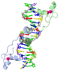
===DNA RECOGNITION BY GAL4: STRUCTURE OF A PROTEIN/DNA COMPLEX=== | ===DNA RECOGNITION BY GAL4: STRUCTURE OF A PROTEIN/DNA COMPLEX=== | ||
| - | {{ | + | {{A specific DNA complex of the 65-residue, N-terminal fragment of the yeast transcriptional activator, GAL4, has been analysed at 2.7 A resolution by X-ray crystallography. The protein binds as a dimer to a symmetrical 17-base-pair sequence. A small, Zn(2+)-containing domain recognizes a conserved CCG triplet at each end of the site through direct contacts with the major groove. A short coiled-coil dimerization element imposes 2-fold symmetry. A segment of extended polypeptide chain links the metal-binding module to the dimerization element and specifies the length of the site. The relatively open structure of the complex would allow another protein to bind coordinately with GAL4.}} |
==About this Structure== | ==About this Structure== | ||
Current revision
| |||||||||
| 1d66, resolution 2.70Å () | |||||||||
|---|---|---|---|---|---|---|---|---|---|
| Ligands: | |||||||||
| |||||||||
| |||||||||
| Resources: | FirstGlance, OCA, RCSB, PDBsum | ||||||||
| Coordinates: | save as pdb, mmCIF, xml | ||||||||
Contents |
DNA RECOGNITION BY GAL4: STRUCTURE OF A PROTEIN/DNA COMPLEX
{{A specific DNA complex of the 65-residue, N-terminal fragment of the yeast transcriptional activator, GAL4, has been analysed at 2.7 A resolution by X-ray crystallography. The protein binds as a dimer to a symmetrical 17-base-pair sequence. A small, Zn(2+)-containing domain recognizes a conserved CCG triplet at each end of the site through direct contacts with the major groove. A short coiled-coil dimerization element imposes 2-fold symmetry. A segment of extended polypeptide chain links the metal-binding module to the dimerization element and specifies the length of the site. The relatively open structure of the complex would allow another protein to bind coordinately with GAL4.}}
About this Structure
1d66 is a 4 chain structure with sequence from Saccharomyces cerevisiae. Full crystallographic information is available from OCA.
See Also
- Gal4
- Hydrogen in macromolecular models
- User:Eric Martz/Introduction to Structural Bioinformatics I
- User:Eric Martz/Introduction to Structural Bioinformatics I%2C 2013
- User:Wayne Decatur/Biochem642 Molecular Visualization 2010 Fall Sessions
- User:Wayne Decatur/Biochem642 Molecular Visualization Sessions
Reference
- Marmorstein R, Carey M, Ptashne M, Harrison SC. DNA recognition by GAL4: structure of a protein-DNA complex. Nature. 1992 Apr 2;356(6368):408-14. PMID:1557122 doi:http://dx.doi.org/10.1038/356408a0


