Sandbox UC 11
From Proteopedia
(Difference between revisions)
| (6 intermediate revisions not shown.) | |||
| Line 1: | Line 1: | ||
| - | {{STRUCTURE_1mnm}} | ||
<StructureSection load='1mnm' size='350' side='right' caption='YEAST MATALPHA2/MCM1/DNA TERNARY TRANSCRIPTION COMPLEX CRYSTAL STRUCTURE (PDB entry [[1mnm]])' scene=''> | <StructureSection load='1mnm' size='350' side='right' caption='YEAST MATALPHA2/MCM1/DNA TERNARY TRANSCRIPTION COMPLEX CRYSTAL STRUCTURE (PDB entry [[1mnm]])' scene=''> | ||
The structure of a complex containing the homeodomain repressor protein MATalpha2 and the MADS-box transcription factor '''MCM1''' bound to DNA has been determined by X-ray crystallography at 2.25 A resolution. | The structure of a complex containing the homeodomain repressor protein MATalpha2 and the MADS-box transcription factor '''MCM1''' bound to DNA has been determined by X-ray crystallography at 2.25 A resolution. | ||
| - | + | [[MtrF]] is a cell surface cytochrome on the Gram-negative bacteria known as ''Shewanella oneidensis''.<ref name="mtrf"> PMID:11418600</ref> MtrF is involved with shuttling electrons across its (''S. oneidensis'') outer surface. MtrF has several homologues, MtrC and the protein OmcA. These three different proteins are thought to be replaceable with one another in deletion mutation experiments.<ref name= "mtrf" /> | |
| + | __TOC__ | ||
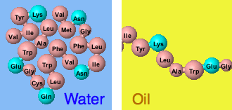
[[Image:MW_Folding_Simulations.gif]] | [[Image:MW_Folding_Simulations.gif]] | ||
| Line 9: | Line 9: | ||
<scene name='Sandbox_UC_11/Structura_secundaria/1'>TextToBeDisplayed</scene> | <scene name='Sandbox_UC_11/Structura_secundaria/1'>TextToBeDisplayed</scene> | ||
| + | ==Structure== | ||
Through the structure of this oligomer that was obtained by crystallography, we can see that HDAC9 is kept <scene name='Sandbox_Reserved_1/Binding/3'>relatively far from the genomic DNA </scene> even after MEF2 binding to the chromosome. We can also easily observe a very well <scene name='Sandbox_Reserved_1/Conservation/7'>evolutionary conserved region</scene> near the DNA binding site of MEF2. | Through the structure of this oligomer that was obtained by crystallography, we can see that HDAC9 is kept <scene name='Sandbox_Reserved_1/Binding/3'>relatively far from the genomic DNA </scene> even after MEF2 binding to the chromosome. We can also easily observe a very well <scene name='Sandbox_Reserved_1/Conservation/7'>evolutionary conserved region</scene> near the DNA binding site of MEF2. | ||
{{Template:ColorKey_Amino2CarboxyRainbow}} | {{Template:ColorKey_Amino2CarboxyRainbow}} | ||
| + | </StructureSection> | ||
| + | ==Directed evolution and Colicin7/Immunity-proteins complexes<ref>PMID:19749752</ref>== | ||
| + | <StructureSection load='3gkl' size='500' frame='true' align='right' scene='3gkl/Al/1' > | ||
| + | {{Clear}} | ||
| + | <scene name='3gkl/Align/2'>Structural alignment</scene> of the immunity protein 9 (Im9, [[1bxi]], <span style="color:yellow;background-color:black;font-weight:bold;">colored yellow</span>), <span style="color:lime;background-color:black;font-weight:bold;">evolved variant R12-2 (lime)</span>, and <font color='blue'><b>immunity protein 7 (Im7, colored blue</b></font>, [[7cei]]) reveals their structural identity. However, when the immunity proteins-bound <scene name='3gkl/Align/3'>colicins within their complexes were aligned</scene>, they demonstrate somewhat different picture. | ||
| + | {{Clear}} | ||
<quiz display= simple> | <quiz display= simple> | ||
| Line 22: | Line 29: | ||
</ Quiz> | </ Quiz> | ||
| - | ==Directed evolution and Colicin7/Immunity-proteins complexes<ref>PMID:19749752</ref>== | ||
| - | <StructureSection load='3gkl' size='500' frame='true' align='right' scene='3gkl/Al/1' > | ||
| - | Iterative rounds of random mutagenesis and selection of <span style="color:yellow;background-color:black;font-weight:bold;">immunity protein 9 (colored yellow)</span> toward higher affinity for ColE7, and selectivity (against ColE9 inhibition), led to significant increase in affinity and selectivity. Several evolved variants were obtained. The crystal structures of the two final generation <scene name='3gkl/Al/3'>variants</scene> <span style="color:lime;background-color:black;font-weight:bold;">R12-2</span> ('''3gkl'''; T20A, N24D, T27A, S28T, V34D, V37J, E41G, and K57E) and <font color='darkred'><b>R12-13</b></font> ([[3gjn]]; N24D, D25E, T27A, S28T, V34D, V37J, and Y55W) in complex with ColE7 were solved. | ||
| - | |||
| - | {{Clear}} | ||
| - | <scene name='3gkl/Align/2'>Structural alignment</scene> of the immunity protein 9 (Im9, [[1bxi]], <span style="color:yellow;background-color:black;font-weight:bold;">colored yellow</span>), <span style="color:lime;background-color:black;font-weight:bold;">evolved variant R12-2 (lime)</span>, and <font color='blue'><b>immunity protein 7 (Im7, colored blue</b></font>, [[7cei]]) reveals their structural identity. However, when the immunity proteins-bound <scene name='3gkl/Align/3'>colicins within their complexes were aligned</scene>, they demonstrate somewhat different picture. The Im9 and Im7 are differ more in their binding configurations (19°, with Tyr54-Tyr55 as the pivot), while the variant R12-2 is in an intermediate configuration between Im9 and Im7. Of note, in the variant R12-2 (3gkl) and Im9 ([[1bxi]]) there are Tyr54 and Tyr55, while in the Im7 ([[7cei]]) Tyr55 and Tyr56 are homologous to them. The most <scene name='3gkl/Align/4'>prominent differences</scene> are in the loop between helices α1 and α2 in Im9 (yellow, labeled in black) and <span style="color:lime;background-color:black;font-weight:bold;">evolved variant R12-2 (lime, labeled in black)</span>. This loop consists of three mutations: N24D, T27A, and S28T in variant R12-2. We can see the deviations in the relative position of helices α1 and α2, in the loop's backbone and in the side chains of residues 24, 26 and 28. | ||
| - | |||
| - | {{Clear}} | ||
MCM1 bound to DNA <ref> '''MCM1''' bound to DNA </ref> | MCM1 bound to DNA <ref> '''MCM1''' bound to DNA </ref> | ||
</StructureSection> | </StructureSection> | ||
<references/> | <references/> | ||
| + | |||
| + | {{STRUCTURE_1mnm}} | ||
==Additional Literature== | ==Additional Literature== | ||
Current revision
| |||||||||||
Directed evolution and Colicin7/Immunity-proteins complexes[2]
| |||||||||||
- ↑ 1.0 1.1 Fotinou C, Emsley P, Black I, Ando H, Ishida H, Kiso M, Sinha KA, Fairweather NF, Isaacs NW. The crystal structure of tetanus toxin Hc fragment complexed with a synthetic GT1b analogue suggests cross-linking between ganglioside receptors and the toxin. J Biol Chem. 2001 Aug 24;276(34):32274-81. Epub 2001 Jun 19. PMID:11418600 doi:10.1074/jbc.M103285200
- ↑ Levin KB, Dym O, Albeck S, Magdassi S, Keeble AH, Kleanthous C, Tawfik DS. Following evolutionary paths to protein-protein interactions with high affinity and selectivity. Nat Struct Mol Biol. 2009 Oct;16(10):1049-55. Epub 2009 Sep 13. PMID:19749752 doi:10.1038/nsmb.1670
| |||||||||
| 1mnm, resolution 2.25Å () | |||||||||
|---|---|---|---|---|---|---|---|---|---|
| |||||||||
| |||||||||
| Resources: | FirstGlance, OCA, RCSB, PDBsum | ||||||||
| Coordinates: | save as pdb, mmCIF, xml | ||||||||
Additional Literature
- Kleanthous C, Kuhlmann UC, Pommer AJ, Ferguson N, Radford SE, Moore GR, James R, Hemmings AM. Structural and mechanistic basis of immunity toward endonuclease colicins. Nat Struct Biol. 1999 Mar;6(3):243-52. PMID:10074943 doi:http://dx.doi.org/10.1038/6683
- Levin KB, Dym O, Albeck S, Magdassi S, Keeble AH, Kleanthous C, Tawfik DS. Following evolutionary paths to protein-protein interactions with high affinity and selectivity. Nat Struct Mol Biol. 2009 Oct;16(10):1049-55. Epub 2009 Sep 13. PMID:19749752 doi:10.1038/nsmb.1670
External Resources
- Yahoo news coverage of luciferase studies funded research for military applications
- Luciferase at Wikipedia
- Luciferase: June 2006 Molecule of the Month as part of the series of tutorials that are at the RCSB Protein Data Bank and written by
- Lipase, the main article in Proteopedia.
- Molecular Playground/Pancreatic Lipase
- Lipase in Wikipedia


