Sandbox Reserved 716
From Proteopedia
m (→'''3D structure''') |
|||
| (67 intermediate revisions not shown.) | |||
| Line 2: | Line 2: | ||
{{Sandbox_ESBS_2012}} | {{Sandbox_ESBS_2012}} | ||
<!-- PLEASE ADD YOUR CONTENT BELOW HERE --> | <!-- PLEASE ADD YOUR CONTENT BELOW HERE --> | ||
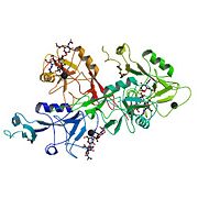
| - | <Structure load='1g1r' size='400' frame='true' align='right' caption='Crystal structure of P-selectin lectin/EGF domains complexed with SLeX' scene='Insert optional scene name here' /> | ||
| + | |||
| + | <big>'''Crystal structure of P-selectin lectin/EGF domains complexed with SLeX'''</big> | ||
| + | {{STRUCTURE_1g1r| PDB=1g1r | SCENE= }} | ||
| + | |||
| + | == '''Introduction''' == | ||
| + | |||
| + | [[Image:1g1r bio r 500.jpg |thumb|left|1g1r]] | ||
| + | |||
| + | |||
| + | |||
| + | |||
| + | Selectins are proteins that are included in a family of cell adhesion receptor involved in the leukocyte extravasation. There are 3 kinds of selectins : | ||
| + | '''E selectin''' localized in endothelial cells, '''L selectin''' found in leukocytes, and '''P selectins''' in platelets and endothelial cells. | ||
| + | |||
| + | In this page we will focus only on P-Selectin. | ||
| + | |||
| + | When the P-selectin is activated by molecules like histamine or thrombin, it is able to bind myeloid cells and some T cells. | ||
| - | '''Crystal structure of P-selectin lectin/EGF domains complexed with SLeX''' | ||
| Line 14: | Line 29: | ||
| - | == '''Introduction''' == | ||
| - | Selectins are proteins that are include in a family of cell adhesion receptor involved in the leukocyte extravasation. There are 3 kinds of selectins : | ||
| - | '''E selectin''' localized in endothelial cells, '''L selectin''' found in leukocytes, and '''P selectins''' in platelets and endothelial cells. | ||
| - | In this page we will be focused only on P-Selectin. | ||
| - | |||
== '''3D structure''' == | == '''3D structure''' == | ||
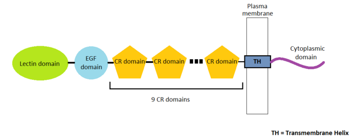
| - | P-Selectin is a protein composed | + | <StructureSection load='1g1r' size='400' side='left' caption='Structure of P-selectin lectin/EGF domains complexed with SLeX (PDB entry [[1g1r]])' scene=''>P-Selectin is a protein composed of 4 different chains A, B, C and D containing 162 amino-acids residues. There are many domains in this protein. Three types of domains represent the extracellular part of the protein : the '''EGF domain''', the '''lectin domain''' and nine '''consensus repeat (CR) domains'''. Finally, we find a transmembrane helix and a cytoplasmic domain.<ref>PMID 11081633</ref> |
| - | EGF domain | + | In this page we will only detail the EGF domain and the lectin domain. |
| - | [[Image: | + | [[Image:Selectin_Structure.png|700px|thumb|left|Scheme of P-selectin strucuture]] |
| - | Lectin domains (also known as C-type lectin domains) are classified in 17 groups (from I to XVII). P-Selectin lectin bellow to the group IV. | ||
| - | [[Image:368px-Pselectin.PNG|200px|center|thumb| P selectin lectin bound to sugar]] | ||
| - | == ''' | + | |
| + | |||
| + | |||
| + | |||
| + | |||
| + | |||
| + | |||
| + | |||
| + | |||
| + | |||
| + | |||
| + | |||
| + | |||
| + | |||
| + | |||
| + | |||
| + | |||
| + | |||
| + | |||
| + | |||
| + | |||
| + | |||
| + | |||
| + | |||
| + | |||
| + | |||
| + | |||
| + | |||
| + | |||
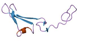
| + | [[Image:PDB 1hre EBI2.jpg|300px|right|thumb| EGF domain]] <scene name='Sandbox_Reserved_716/Egf_domain/1'>EGF domain</scene> (Epidermal Growth Factor) contains 30 to 40 amino-acids residues. This domain is composed of 6 cysteine residues which can form 3 disulfide bonds. EGF domain is formed with two-stranded β-sheet followed by a loop to a short C-terminal two-stranded β-sheet. These two β-sheets are usually denoted as the major (N-terminal) and minor (C-terminal) sheets. | ||
| + | |||
| + | EGF domain seems to be useful for three kind of interactions : | ||
| + | |||
| + | - '''ligand recognition''' : The main ligand for P-selectin is P-selectin glycoprotein ligand-1 (PSGL-1) | ||
| + | |||
| + | - '''cell adhesion''' : adhesion of the leukocyte to the vessel membrane | ||
| + | |||
| + | - '''protein-protein interactions'''<ref>PMID 7508943</ref> | ||
| + | |||
| + | |||
| + | |||
| + | |||
| + | |||
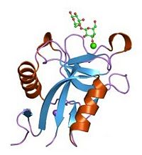
| + | [[Image:Lectin domain.jpg|200px|left|thumb| Lectin domain bound to sugar]] | ||
| + | |||
| + | |||
| + | Lectin domains (also known as C-type lectin domains) are classified in 17 groups (from I to XVII). P-Selectin lectin domain belongs to the group IV. | ||
| + | |||
| + | <scene name='Sandbox_Reserved_716/Lectin_domain/2'>P-selectin lectin domain</scene> is composed by 118 amino-acids residues and is a succession of β-sheets and helix. | ||
| + | |||
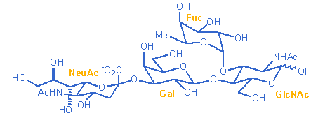
| + | It binds (via an extended site of the domain) a ligand : '''the carbohydrate sialyl LewisX (SLeX)''' with low affinity. However, the affinity between the ligand and the selectins is actually quite high. A theory exists and may explain why lectin domain may recognize SLeX (or related carbohydrate structures on this ligand) with a higher affinity. Indeed EGF domain may alter the conformation of the lectin domain so that its affinity for the ligand is increased. | ||
| + | [[Image:Slex.png|500px|left|thumb| SLeX]] | ||
| + | |||
| + | |||
| + | |||
| + | |||
| + | |||
| + | |||
| + | Sialyl LewisX is a tetrasaccharide carbohydrate that is usually attached to O-glycans on the surface of cells. It plays a role in cell-to-cell recognition processes. It binds making hydrogene bonds with Tyr48, Asn82, Glu92, Tyr94, Arg97, Ser99, Asn105, Glu107 of P-selectin. | ||
| + | |||
| + | </StructureSection> | ||
| + | |||
| + | == '''Different roles of P-selectin''' == | ||
=== '''Role in leukocyte extravasation''' === | === '''Role in leukocyte extravasation''' === | ||
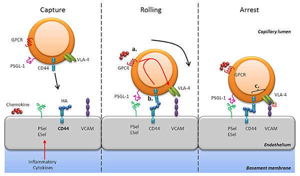
| - | Leukocyte extravasation is the movement of leukocytes out of the circulatory system. First, the | + | Leukocyte extravasation is the movement of leukocytes out of the circulatory system during an infection. The inflammation is a natural reaction of defense caused by tissular damages. The recruitement of leukocytes on the inflammation site is a process in four steps. First, the leukocyte is attracted by cytokines, secreted near the site of infection (these cytokines are also able to activate P-selectins). This is the chemoattraction. Then, this leukocyte begin rolling along the inner surface of the vessel, '''binding on selectin molecules'''. The third step is the tight adhesion, the leukocyte immobilizates itself, thanks to the integrin molecules. And finally, he passes through gaps between endothelial cells. This last step is not shown in the following drawing. By this mecanism, the leukocyte arrives on the site of infection to neutralize the infection agent.<ref>[http://www.ircm.qc.ca/LARECHERCHE/axes/Biologie/Chimie/Documents/Guindon_ProjetChimieCarbo.pdf Chimie des carbohydrates : Mimétiques du sialyl Lewis X] par Benoit Cardinal-David et Daniel Charpentier</ref> |
| + | [[Image:Receptors.jpg|600px|thumb|center|Leukocyte extravasation receptors and role of P-selectin in this mechanism]] | ||
=== '''Role in platelets recruitment''' === | === '''Role in platelets recruitment''' === | ||
| + | |||
| + | At areas of vascular injury, P-selectin recruits and aggregates platelets<ref>PMID 16845394</ref> through platelet-fibrin and platelet-platelet binding. In quiescent platelet, P-selectin is located on the inner wall of α-granules. The activation of platelets is caused by thrombin, Type II collagen and ADP and results in "membrane flipping" where the platelet releases α- and dense granules and the inner walls of the granules are exposed on the outside of the cell. | ||
=== '''Role in cancer''' === | === '''Role in cancer''' === | ||
| + | P-selectin also has a functional role in formation of tumor. P-selectin is expressed on the surface of both stimulated endothelial cell and activated platelet. It helps cancer cells invade into bloodstream for metastasis. Moreover, platelet facilitates tumor metastasis by forming complexes with tumor cells and leukocytes. In vivo mice experiment showed that reduction in circulating platelets could reduce cancer metastasis.<ref>PMID 16648968</ref> So we can understand the importance of development of new compounds that target P-selectin for cancer therapy. | ||
| + | == '''External resources''' == | ||
| + | Protein Data Bank file [http://www.rcsb.org/pdb/explore/explore.do?structureId=1G1R 1G1R] | ||
== '''References''' == | == '''References''' == | ||
| - | + | <references/> | |
| - | + | ||
| - | + | ||
Current revision
Crystal structure of P-selectin lectin/EGF domains complexed with SLeX
| |||||||||
| 1g1r, resolution 3.40Å () | |||||||||
|---|---|---|---|---|---|---|---|---|---|
| Ligands: | , , , , | ||||||||
| Non-Standard Residues: | |||||||||
| Related: | 1g1q, 1g1s, 1g1t | ||||||||
| |||||||||
| |||||||||
| Resources: | FirstGlance, OCA, RCSB, PDBsum | ||||||||
| Coordinates: | save as pdb, mmCIF, xml | ||||||||
Contents |
Introduction
Selectins are proteins that are included in a family of cell adhesion receptor involved in the leukocyte extravasation. There are 3 kinds of selectins :
E selectin localized in endothelial cells, L selectin found in leukocytes, and P selectins in platelets and endothelial cells.
In this page we will focus only on P-Selectin.
When the P-selectin is activated by molecules like histamine or thrombin, it is able to bind myeloid cells and some T cells.
3D structure
| |||||||||||
Different roles of P-selectin
Role in leukocyte extravasation
Leukocyte extravasation is the movement of leukocytes out of the circulatory system during an infection. The inflammation is a natural reaction of defense caused by tissular damages. The recruitement of leukocytes on the inflammation site is a process in four steps. First, the leukocyte is attracted by cytokines, secreted near the site of infection (these cytokines are also able to activate P-selectins). This is the chemoattraction. Then, this leukocyte begin rolling along the inner surface of the vessel, binding on selectin molecules. The third step is the tight adhesion, the leukocyte immobilizates itself, thanks to the integrin molecules. And finally, he passes through gaps between endothelial cells. This last step is not shown in the following drawing. By this mecanism, the leukocyte arrives on the site of infection to neutralize the infection agent.[3]
Role in platelets recruitment
At areas of vascular injury, P-selectin recruits and aggregates platelets[4] through platelet-fibrin and platelet-platelet binding. In quiescent platelet, P-selectin is located on the inner wall of α-granules. The activation of platelets is caused by thrombin, Type II collagen and ADP and results in "membrane flipping" where the platelet releases α- and dense granules and the inner walls of the granules are exposed on the outside of the cell.
Role in cancer
P-selectin also has a functional role in formation of tumor. P-selectin is expressed on the surface of both stimulated endothelial cell and activated platelet. It helps cancer cells invade into bloodstream for metastasis. Moreover, platelet facilitates tumor metastasis by forming complexes with tumor cells and leukocytes. In vivo mice experiment showed that reduction in circulating platelets could reduce cancer metastasis.[5] So we can understand the importance of development of new compounds that target P-selectin for cancer therapy.
External resources
Protein Data Bank file 1G1R
References
- ↑ Somers WS, Tang J, Shaw GD, Camphausen RT. Insights into the molecular basis of leukocyte tethering and rolling revealed by structures of P- and E-selectin bound to SLe(X) and PSGL-1. Cell. 2000 Oct 27;103(3):467-79. PMID:11081633
- ↑ Kansas GS, Saunders KB, Ley K, Zakrzewicz A, Gibson RM, Furie BC, Furie B, Tedder TF. A role for the epidermal growth factor-like domain of P-selectin in ligand recognition and cell adhesion. J Cell Biol. 1994 Feb;124(4):609-18. PMID:7508943
- ↑ Chimie des carbohydrates : Mimétiques du sialyl Lewis X par Benoit Cardinal-David et Daniel Charpentier
- ↑ Phan UT, Waldron TT, Springer TA. Remodeling of the lectin-EGF-like domain interface in P- and L-selectin increases adhesiveness and shear resistance under hydrodynamic force. Nat Immunol. 2006 Aug;7(8):883-9. Epub 2006 Jul 16. PMID:16845394 doi:10.1038/ni1366
- ↑ Chen M, Geng JG. P-selectin mediates adhesion of leukocytes, platelets, and cancer cells in inflammation, thrombosis, and cancer growth and metastasis. Arch Immunol Ther Exp (Warsz). 2006 Mar-Apr;54(2):75-84. Epub 2006 Mar 24. PMID:16648968 doi:10.1007/s00005-006-0010-6
Proteopedia Page Contributors and Editors
Delphine Trelat, Cécile Ehrhardt

