This old version of Proteopedia is provided for student assignments while the new version is undergoing repairs. Content and edits done in this old version of Proteopedia after March 1, 2026 will eventually be lost when it is retired in about June of 2026.
Apply for new accounts at the new Proteopedia. Your logins will work in both the old and new versions.
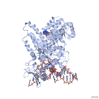
1gm5
From Proteopedia
(Difference between revisions)
| (7 intermediate revisions not shown.) | |||
| Line 1: | Line 1: | ||
| - | {{STRUCTURE_1gm5| PDB=1gm5 | SCENE= }} | ||
| - | ===STRUCTURE OF RECG BOUND TO THREE-WAY DNA JUNCTION=== | ||
| - | {{ABSTRACT_PUBMED_11595187}} | ||
| - | == | + | ==Structure of RecG bound to three-way DNA junction== |
| - | [[1gm5]] is a 4 chain structure with sequence from [ | + | <StructureSection load='1gm5' size='340' side='right'caption='[[1gm5]], [[Resolution|resolution]] 3.24Å' scene=''> |
| + | == Structural highlights == | ||
| + | <table><tr><td colspan='2'>[[1gm5]] is a 4 chain structure with sequence from [https://en.wikipedia.org/wiki/Thermotoga_maritima Thermotoga maritima]. Full crystallographic information is available from [http://oca.weizmann.ac.il/oca-bin/ocashort?id=1GM5 OCA]. For a <b>guided tour on the structure components</b> use [https://proteopedia.org/fgij/fg.htm?mol=1GM5 FirstGlance]. <br> | ||
| + | </td></tr><tr id='method'><td class="sblockLbl"><b>[[Empirical_models|Method:]]</b></td><td class="sblockDat" id="methodDat">X-ray diffraction, [[Resolution|Resolution]] 3.24Å</td></tr> | ||
| + | <tr id='ligand'><td class="sblockLbl"><b>[[Ligand|Ligands:]]</b></td><td class="sblockDat" id="ligandDat"><scene name='pdbligand=ADP:ADENOSINE-5-DIPHOSPHATE'>ADP</scene>, <scene name='pdbligand=MG:MAGNESIUM+ION'>MG</scene></td></tr> | ||
| + | <tr id='resources'><td class="sblockLbl"><b>Resources:</b></td><td class="sblockDat"><span class='plainlinks'>[https://proteopedia.org/fgij/fg.htm?mol=1gm5 FirstGlance], [http://oca.weizmann.ac.il/oca-bin/ocaids?id=1gm5 OCA], [https://pdbe.org/1gm5 PDBe], [https://www.rcsb.org/pdb/explore.do?structureId=1gm5 RCSB], [https://www.ebi.ac.uk/pdbsum/1gm5 PDBsum], [https://prosat.h-its.org/prosat/prosatexe?pdbcode=1gm5 ProSAT]</span></td></tr> | ||
| + | </table> | ||
| + | == Function == | ||
| + | [https://www.uniprot.org/uniprot/Q9WY48_THEMA Q9WY48_THEMA] | ||
| + | == Evolutionary Conservation == | ||
| + | [[Image:Consurf_key_small.gif|200px|right]] | ||
| + | Check<jmol> | ||
| + | <jmolCheckbox> | ||
| + | <scriptWhenChecked>; select protein; define ~consurf_to_do selected; consurf_initial_scene = true; script "/wiki/ConSurf/gm/1gm5_consurf.spt"</scriptWhenChecked> | ||
| + | <scriptWhenUnchecked>script /wiki/extensions/Proteopedia/spt/initialview01.spt</scriptWhenUnchecked> | ||
| + | <text>to colour the structure by Evolutionary Conservation</text> | ||
| + | </jmolCheckbox> | ||
| + | </jmol>, as determined by [http://consurfdb.tau.ac.il/ ConSurfDB]. You may read the [[Conservation%2C_Evolutionary|explanation]] of the method and the full data available from [http://bental.tau.ac.il/new_ConSurfDB/main_output.php?pdb_ID=1gm5 ConSurf]. | ||
| + | <div style="clear:both"></div> | ||
==See Also== | ==See Also== | ||
| + | *[[Helicase 3D structures|Helicase 3D structures]] | ||
| + | *[[RecG|RecG]] | ||
*[[RecG Bound to Three-Way DNA Junction|RecG Bound to Three-Way DNA Junction]] | *[[RecG Bound to Three-Way DNA Junction|RecG Bound to Three-Way DNA Junction]] | ||
| - | + | __TOC__ | |
| - | + | </StructureSection> | |
| - | < | + | [[Category: Large Structures]] |
[[Category: Thermotoga maritima]] | [[Category: Thermotoga maritima]] | ||
| - | [[Category: Scaife | + | [[Category: Scaife S]] |
| - | [[Category: Singleton | + | [[Category: Singleton MR]] |
| - | [[Category: Wigley | + | [[Category: Wigley DB]] |
| - | + | ||
| - | + | ||
Current revision
Structure of RecG bound to three-way DNA junction
| |||||||||||