1p8x
From Proteopedia
(Difference between revisions)
| (5 intermediate revisions not shown.) | |||
| Line 1: | Line 1: | ||
| - | {{STRUCTURE_1p8x| PDB=1p8x | SCENE= }} | ||
| - | ===The Calcium-Activated C-terminal half of gelsolin=== | ||
| - | {{ABSTRACT_PUBMED_14527664}} | ||
| - | == | + | ==The Calcium-Activated C-terminal half of gelsolin== |
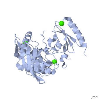
| - | [[http://www.uniprot.org/uniprot/GELS_HUMAN GELS_HUMAN | + | <StructureSection load='1p8x' size='340' side='right'caption='[[1p8x]], [[Resolution|resolution]] 2.00Å' scene=''> |
| + | == Structural highlights == | ||
| + | <table><tr><td colspan='2'>[[1p8x]] is a 3 chain structure with sequence from [https://en.wikipedia.org/wiki/Homo_sapiens Homo sapiens]. Full crystallographic information is available from [http://oca.weizmann.ac.il/oca-bin/ocashort?id=1P8X OCA]. For a <b>guided tour on the structure components</b> use [https://proteopedia.org/fgij/fg.htm?mol=1P8X FirstGlance]. <br> | ||
| + | </td></tr><tr id='method'><td class="sblockLbl"><b>[[Empirical_models|Method:]]</b></td><td class="sblockDat" id="methodDat">X-ray diffraction, [[Resolution|Resolution]] 2Å</td></tr> | ||
| + | <tr id='ligand'><td class="sblockLbl"><b>[[Ligand|Ligands:]]</b></td><td class="sblockDat" id="ligandDat"><scene name='pdbligand=CA:CALCIUM+ION'>CA</scene></td></tr> | ||
| + | <tr id='resources'><td class="sblockLbl"><b>Resources:</b></td><td class="sblockDat"><span class='plainlinks'>[https://proteopedia.org/fgij/fg.htm?mol=1p8x FirstGlance], [http://oca.weizmann.ac.il/oca-bin/ocaids?id=1p8x OCA], [https://pdbe.org/1p8x PDBe], [https://www.rcsb.org/pdb/explore.do?structureId=1p8x RCSB], [https://www.ebi.ac.uk/pdbsum/1p8x PDBsum], [https://prosat.h-its.org/prosat/prosatexe?pdbcode=1p8x ProSAT]</span></td></tr> | ||
| + | </table> | ||
| + | == Disease == | ||
| + | [https://www.uniprot.org/uniprot/GELS_HUMAN GELS_HUMAN] Defects in GSN are the cause of amyloidosis type 5 (AMYL5) [MIM:[https://omim.org/entry/105120 105120]; also known as familial amyloidosis Finnish type. AMYL5 is a hereditary generalized amyloidosis due to gelsolin amyloid deposition. It is typically characterized by cranial neuropathy and lattice corneal dystrophy. Most patients have modest involvement of internal organs, but severe systemic disease can develop in some individuals causing peripheral polyneuropathy, amyloid cardiomyopathy, and nephrotic syndrome leading to renal failure.<ref>PMID:2157434</ref> <ref>PMID:2153578</ref> <ref>PMID:2176481</ref> <ref>PMID:1338910</ref> | ||
| + | == Function == | ||
| + | [https://www.uniprot.org/uniprot/GELS_HUMAN GELS_HUMAN] Calcium-regulated, actin-modulating protein that binds to the plus (or barbed) ends of actin monomers or filaments, preventing monomer exchange (end-blocking or capping). It can promote the assembly of monomers into filaments (nucleation) as well as sever filaments already formed. Plays a role in ciliogenesis.<ref>PMID:20393563</ref> | ||
| + | == Evolutionary Conservation == | ||
| + | [[Image:Consurf_key_small.gif|200px|right]] | ||
| + | Check<jmol> | ||
| + | <jmolCheckbox> | ||
| + | <scriptWhenChecked>; select protein; define ~consurf_to_do selected; consurf_initial_scene = true; script "/wiki/ConSurf/p8/1p8x_consurf.spt"</scriptWhenChecked> | ||
| + | <scriptWhenUnchecked>script /wiki/extensions/Proteopedia/spt/initialview01.spt</scriptWhenUnchecked> | ||
| + | <text>to colour the structure by Evolutionary Conservation</text> | ||
| + | </jmolCheckbox> | ||
| + | </jmol>, as determined by [http://consurfdb.tau.ac.il/ ConSurfDB]. You may read the [[Conservation%2C_Evolutionary|explanation]] of the method and the full data available from [http://bental.tau.ac.il/new_ConSurfDB/main_output.php?pdb_ID=1p8x ConSurf]. | ||
| + | <div style="clear:both"></div> | ||
| + | <div style="background-color:#fffaf0;"> | ||
| + | == Publication Abstract from PubMed == | ||
| + | Gelsolin requires activation to carry out its severing and capping activities on F-actin. Here, we present the structure of the isolated C-terminal half of gelsolin (G4-G6) at 2.0 A resolution in the presence of Ca(2+) ions. This structure completes a triptych of the states of activation of G4-G6 that illuminates its role in the function of gelsolin. Activated G4-G6 displays an open conformation, with the actin-binding site on G4 fully exposed and all three type-2 Ca(2+) sites occupied. Neither actin nor the type-l Ca(2+), which normally is sandwiched between actin and G4, is required to achieve this conformation. | ||
| - | + | Activation in isolation: exposure of the actin-binding site in the C-terminal half of gelsolin does not require actin.,Narayan K, Chumnarnsilpa S, Choe H, Irobi E, Urosev D, Lindberg U, Schutt CE, Burtnick LD, Robinson RC FEBS Lett. 2003 Sep 25;552(2-3):82-5. PMID:14527664<ref>PMID:14527664</ref> | |
| - | + | ||
| - | + | From MEDLINE®/PubMed®, a database of the U.S. National Library of Medicine.<br> | |
| - | + | </div> | |
| + | <div class="pdbe-citations 1p8x" style="background-color:#fffaf0;"></div> | ||
==See Also== | ==See Also== | ||
| - | *[[Gelsolin|Gelsolin]] | + | *[[Gelsolin 3D structures|Gelsolin 3D structures]] |
| - | + | == References == | |
| - | == | + | <references/> |
| - | + | __TOC__ | |
| + | </StructureSection> | ||
[[Category: Homo sapiens]] | [[Category: Homo sapiens]] | ||
| - | [[Category: | + | [[Category: Large Structures]] |
| - | [[Category: | + | [[Category: Burtnick LD]] |
| - | [[Category: | + | [[Category: Narayan K]] |
| - | [[Category: | + | [[Category: Robinson RC]] |
| - | + | ||
Current revision
The Calcium-Activated C-terminal half of gelsolin
| |||||||||||