| Important biological processes, such as synaptic transmission and cellular trafficking in Eukaryotes require SNARE proteins that are thought to play a crucial role in membrane fusion.[1][2][3][4] To connect membranes and allow their fusion, SNARE (soluble N-ethylmaleimide-sensitive-factor attachment protein receptor) proteins assemble into a core-complex of four parallel helices.[5][6][7] The SNARE complex assembly is mediated by a conserved SNARE motif consisting of 60-70 amino acids. [8] SNAREs can be divided into two categories: the v-(vesicle) SNAREs, which are found in the vesicle membrane and the t-(target) SNAREs, which are anchored in the target membrane.[9]
The best-studied SNAREs are the neuronal and the early endosomal SNARE complexes. The Neuronal SNARE complex mediates exocytosis of synaptic vesicles in the neurons and includes the vesicle protein synaptobrevin (also called VAMP), the membrane proteins SNAP-25 and syntaxin 1.[7] The early endosomal SNARE complex includes syntaxin 6, syntaxin 13, vti1a and VAMP4 and is responsible for homotypic fusion of early endosomes.[10] It has been shown that the crystal structure of the early endosomal SNARE complex resembles that of the neuronal and late endosomal complexes, but differs in surface side-chain interactions.
Membrane fusion mechanism
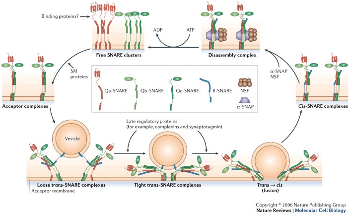
 Membrane fusion mechanism Membrane Fusion requires the assembly of the core complex. Free t-SNAREs that are organized in clusters first assemble into acceptor complexes thanks to SM (Sec1/Munc18-related) proteins. Acceptor complexes can then interact with the v-SNAREs through the N-terminal domain of the SNARE motif. This enables the formation of four-helical trans-complexes, in which only the N-terminal portions of the SNARE motifs are bound. This binding evolves from a loose to a tight state, thus leading to the formation of a fusion pore. During the fusion, the conformation relaxes to a cis-configuration. Cis-complexes dissociate thanks to proteins and cofactors (SNAPs). T- and v-SNAREs can be separated and recycled.[11]
Structure
SNARE domain
The SNARE domain is approximately 60-70 residues long and is located immediately adjacent to a C-terminal transmembrane anchor. It contains a repeating heptad pattern of hydrophobic residues. Their orientation places them in an alpha-helical structure so that all the hydrophobic side chains are located on the same face of the helix.[5][12][7] SNARE domains allow the four SNAREs protein to assemble into parallel four-helix bundles. This parallel arrangement brings the transmembrane anchors and the membranes closer. [13]
“0”-layer
 Topology of the 16 layers of the SNARE complex The centre of the four-helix bundle is constituted of 16 layers. These layers are composed of , which are perpendicular to the axis of the four-helix bundle, except for the central “0”-layer. This last one consists of three glutamine (Q) and one arginine (R) highly conserved residues. Those highly conserved residues have led to a new classification of SNAREs into Q- and R-SNAREs. [14] Almost all membrane fusion reactions require one R-SNARE and three Q-SNAREs: Qa, Qb and Qc.[15][16] In many cases, the R-SNARE is in the vesicle, and the three Q-SNAREs are in the target membrane. For the early endosomal SNARE complex, syntaxin 13, vti1a, syntaxin 6, and VAMP4 were respectively classified as Qa-, Qb-, Qc- and R-SNAREs.
Structure of the early endosomal SNARE complex 2NPS
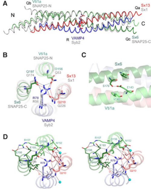
 Crystal structure of the early endosomal SNARE complex. (A) Backbone overlay of the early endosomal SNARE complex (in color) and the neuronal SNARE complex (gray). (B) N- to C-terminal view of the 0 layer containing the unusual aspartate in vti1a. (Color, early endosomal SNARE complex; gray, neuronal SNARE complex.) (C) Residue E143 builds a salt bridge with the residue S179. The residue E143 is also sandwiched by residues V182 and V140, which strengthen polar interactions. In the late endosomal and in the neuronal SNARE complex, this glutamate is conserved and interacts with a conserved arginine that occupies the position equivalent to V182. (D) View of the 0 layer, including hydrogen bonds with surface residues and water molecules.
The early endosomal SNARE complex forms a four-helix bundle with a left-handed superhelical twist. The position of the Qa-, Qb-, Qc-, and R-SNAREs is the same in the neuronal and late endosomal complexes.[7][5] As mentioned above, an unconventional layer of hydrophilic residues – the “0”-layer – constituted of three glutamines and one arginine, characterizes the center of all SNARE complexes. However, in vti1a an aspartate residue substitutes the glutamine. The aspartate occupies the same position as the glutamine in the neuronal and late endosomal complexes, and is involved in a similar organization of hydrogen bonds with the other layer of amino acids. Moreover, in vti1a, of the ‘0' layer also interacts with at the surface of the vti1a helix and with a water molecule. Also, interacts with the and with that in turn interacts with . Concerning , it interacts with . This network of interactions resembles that observed in the neuronal complex.[17] The structure of the other layers is generally similar to that of the neuronal and late endosomal complexes. Indeed, slightly differences can be noted: in layer 6, the Qc-SNARE syntaxin 6 contains a valine, whereas the late endosomal Qc-SNARE syntaxin 8 contains a glutamate, which is involved in interactions with surface residues. Such interactions are absent in the early endosomal and the neuronal complexes.
Regarding the SNARE motifs, they are not only connected by the central interacting layers, but also by surface interactions that often involve side chains with complementary charges. As an example, an interaction between the helices of syntaxin 6 and vti1a involves a . At a similar position in the late endosomal complex, a salt bridge is observed between .[5] It has to be noted that all these side chains are highly conserved between the respective SNAREs of Drosophila and mammals. Many other surface interactions connect the SNARE motifs. However, the role of these surface interactions in the stability of the whole complex is still unknown.
Regulation
In addition to SNARE motifs and membrane anchors, SNARE proteins have autonomously folding N-terminal domains. These autonomously folded domains are able to regulate SNARE assembly. For example, synthaxin 7 has a three-helix-bundle domain, which with the profiling-like domain Ykt6p of yeast can lightly inhibit the SNARE assembly.[18] Sso1p is the yeast ortholog of syntax in. Ssop has also a three-helix-bundle domain that can bind to the SNARE motif. This binding generates a closed conformation that strongly inhibits the entry of Sso1p into SNARE complexes.[19][20][21][22] The main role of these autonomously folded domains is still under research. However, one of the known functions is the recruitment of other trafficking proteins thanks to the open/closed equilibrium of some SNAREs.
SNAREs and pulmonary hypertension
Pulmonary hypertension (PH) corresponds to an increase of blood pressure in the pulmonary artery, pulmonary vein, or pulmonary capillaries leading to shortness of breath, dizziness, fainting, leg swelling and other symptoms. Pulmonary hypertension can be a severe disease, characterized by a decreased exercise tolerance and heart failure. The origin of the disease seems to be a Golgi-dysfunction. Indeed, dysfunction of Golgi tethers, SNAPs and SNAREs apparently leads to pulmonary hypertension.[23]
References
- ↑ Brunger AT. Structure of proteins involved in synaptic vesicle fusion in neurons. Annu Rev Biophys Biomol Struct. 2001;30:157-71. PMID:11340056 doi:http://dx.doi.org/10.1146/annurev.biophys.30.1.157
- ↑ Chen YA, Scheller RH. SNARE-mediated membrane fusion. Nat Rev Mol Cell Biol. 2001 Feb;2(2):98-106. PMID:11252968 doi:http://dx.doi.org/10.1038/35052017
- ↑ Jahn R, Lang T, Sudhof TC. Membrane fusion. Cell. 2003 Feb 21;112(4):519-33. PMID:12600315
- ↑ Rizo J, Sudhof TC. Snares and Munc18 in synaptic vesicle fusion. Nat Rev Neurosci. 2002 Aug;3(8):641-53. PMID:12154365 doi:http://dx.doi.org/10.1038/nrn898
- ↑ 5.0 5.1 5.2 5.3 Antonin W, Fasshauer D, Becker S, Jahn R, Schneider TR. Crystal structure of the endosomal SNARE complex reveals common structural principles of all SNAREs. Nat Struct Biol. 2002 Feb;9(2):107-11. PMID:11786915 doi:http://dx.doi.org/10.1038/nsb746
- ↑ Poirier MA, Xiao W, Macosko JC, Chan C, Shin YK, Bennett MK. The synaptic SNARE complex is a parallel four-stranded helical bundle. Nat Struct Biol. 1998 Sep;5(9):765-9. PMID:9731768 doi:10.1038/1799
- ↑ 7.0 7.1 7.2 7.3 Sutton RB, Fasshauer D, Jahn R, Brunger AT. Crystal structure of a SNARE complex involved in synaptic exocytosis at 2.4 A resolution. Nature. 1998 Sep 24;395(6700):347-53. PMID:9759724 doi:http://dx.doi.org/10.1038/26412
- ↑ Weimbs T, Low SH, Chapin SJ, Mostov KE, Bucher P, Hofmann K. A conserved domain is present in different families of vesicular fusion proteins: a new superfamily. Proc Natl Acad Sci U S A. 1997 Apr 1;94(7):3046-51. PMID:9096343
- ↑ Sollner T, Whiteheart SW, Brunner M, Erdjument-Bromage H, Geromanos S, Tempst P, Rothman JE. SNAP receptors implicated in vesicle targeting and fusion. Nature. 1993 Mar 25;362(6418):318-24. PMID:8455717 doi:http://dx.doi.org/10.1038/362318a0
- ↑ Brandhorst D, Zwilling D, Rizzoli SO, Lippert U, Lang T, Jahn R. Homotypic fusion of early endosomes: SNAREs do not determine fusion specificity. Proc Natl Acad Sci U S A. 2006 Feb 21;103(8):2701-6. Epub 2006 Feb 9. PMID:16469845 doi:http://dx.doi.org/10.1073/pnas.0511138103
- ↑ Jahn R, Scheller RH. SNAREs--engines for membrane fusion. Nat Rev Mol Cell Biol. 2006 Sep;7(9):631-43. Epub 2006 Aug 16. PMID:16912714 doi:http://dx.doi.org/10.1038/nrm2002
- ↑ Poirier MA, Xiao W, Macosko JC, Chan C, Shin YK, Bennett MK. The synaptic SNARE complex is a parallel four-stranded helical bundle. Nat Struct Biol. 1998 Sep;5(9):765-9. PMID:9731768 doi:10.1038/1799
- ↑ Hanson PI, Roth R, Morisaki H, Jahn R, Heuser JE. Structure and conformational changes in NSF and its membrane receptor complexes visualized by quick-freeze/deep-etch electron microscopy. Cell. 1997 Aug 8;90(3):523-35. PMID:9267032
- ↑ Fasshauer D, Sutton RB, Brunger AT, Jahn R. Conserved structural features of the synaptic fusion complex: SNARE proteins reclassified as Q- and R-SNAREs. Proc Natl Acad Sci U S A. 1998 Dec 22;95(26):15781-6. PMID:9861047
- ↑ Bock JB, Matern HT, Peden AA, Scheller RH. A genomic perspective on membrane compartment organization. Nature. 2001 Feb 15;409(6822):839-41. PMID:11237004 doi:http://dx.doi.org/10.1038/35057024
- ↑ McNew JA, Parlati F, Fukuda R, Johnston RJ, Paz K, Paumet F, Sollner TH, Rothman JE. Compartmental specificity of cellular membrane fusion encoded in SNARE proteins. Nature. 2000 Sep 14;407(6801):153-9. PMID:11001046 doi:http://dx.doi.org/10.1038/35025000
- ↑ Ernst JA, Brunger AT. High resolution structure, stability, and synaptotagmin binding of a truncated neuronal SNARE complex. J Biol Chem. 2003 Mar 7;278(10):8630-6. Epub 2002 Dec 20. PMID:12496247 doi:http://dx.doi.org/10.1074/jbc.M211889200
- ↑ Tochio H, Tsui MM, Banfield DK, Zhang M. An autoinhibitory mechanism for nonsyntaxin SNARE proteins revealed by the structure of Ykt6p. Science. 2001 Jul 27;293(5530):698-702. PMID:11474112 doi:10.1126/science.1062950
- ↑ Fiebig KM, Rice LM, Pollock E, Brunger AT. Folding intermediates of SNARE complex assembly. Nat Struct Biol. 1999 Feb;6(2):117-23. PMID:10048921 doi:10.1038/5803
- ↑ Munson M, Chen X, Cocina AE, Schultz SM, Hughson FM. Interactions within the yeast t-SNARE Sso1p that control SNARE complex assembly. Nat Struct Biol. 2000 Oct;7(10):894-902. PMID:11017200 doi:10.1038/79659
- ↑ Munson M, Hughson FM. Conformational regulation of SNARE assembly and disassembly in vivo. J Biol Chem. 2002 Mar 15;277(11):9375-81. Epub 2002 Jan 2. PMID:11777922 doi:http://dx.doi.org/10.1074/jbc.M111729200
- ↑ Nicholson KL, Munson M, Miller RB, Filip TJ, Fairman R, Hughson FM. Regulation of SNARE complex assembly by an N-terminal domain of the t-SNARE Sso1p. Nat Struct Biol. 1998 Sep;5(9):793-802. PMID:9731774 doi:http://dx.doi.org/10.1038/1834
- ↑ Sehgal PB, Mukhopadhyay S. Pulmonary arterial hypertension: a disease of tethers, SNAREs and SNAPs? Am J Physiol Heart Circ Physiol. 2007 Jul;293(1):H77-85. Epub 2007 Apr 6. PMID:17416597 doi:http://dx.doi.org/10.1152/ajpheart.01386.2006
Proteopedia page contributors and editors
Florence HERMAL and Camille ROESCH
|


