We apologize for Proteopedia being slow to respond. For the past two years, a new implementation of Proteopedia has been being built. Soon, it will replace this 18-year old system. All existing content will be moved to the new system at a date that will be announced here.
3l1l
From Proteopedia
(Difference between revisions)
| (4 intermediate revisions not shown.) | |||
| Line 1: | Line 1: | ||
| - | {{STRUCTURE_3l1l| PDB=3l1l | SCENE= }} | ||
| - | ===Structure of Arg-bound Escherichia coli AdiC=== | ||
| - | {{ABSTRACT_PUBMED_20090677}} | ||
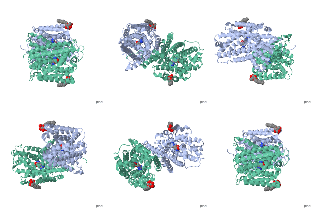
| - | == | + | ==Structure of Arg-bound Escherichia coli AdiC== |
| - | [[http://www.uniprot.org/uniprot/ADIC_ECO57 ADIC_ECO57 | + | <StructureSection load='3l1l' size='340' side='right'caption='[[3l1l]], [[Resolution|resolution]] 3.00Å' scene=''> |
| + | == Structural highlights == | ||
| + | <table><tr><td colspan='2'>[[3l1l]] is a 1 chain structure with sequence from [https://en.wikipedia.org/wiki/Escherichia_coli_O157:H7 Escherichia coli O157:H7]. Full crystallographic information is available from [http://oca.weizmann.ac.il/oca-bin/ocashort?id=3L1L OCA]. For a <b>guided tour on the structure components</b> use [https://proteopedia.org/fgij/fg.htm?mol=3L1L FirstGlance]. <br> | ||
| + | </td></tr><tr id='method'><td class="sblockLbl"><b>[[Empirical_models|Method:]]</b></td><td class="sblockDat" id="methodDat">X-ray diffraction, [[Resolution|Resolution]] 3.002Å</td></tr> | ||
| + | <tr id='ligand'><td class="sblockLbl"><b>[[Ligand|Ligands:]]</b></td><td class="sblockDat" id="ligandDat"><scene name='pdbligand=ARG:ARGININE'>ARG</scene>, <scene name='pdbligand=BNG:B-NONYLGLUCOSIDE'>BNG</scene></td></tr> | ||
| + | <tr id='resources'><td class="sblockLbl"><b>Resources:</b></td><td class="sblockDat"><span class='plainlinks'>[https://proteopedia.org/fgij/fg.htm?mol=3l1l FirstGlance], [http://oca.weizmann.ac.il/oca-bin/ocaids?id=3l1l OCA], [https://pdbe.org/3l1l PDBe], [https://www.rcsb.org/pdb/explore.do?structureId=3l1l RCSB], [https://www.ebi.ac.uk/pdbsum/3l1l PDBsum], [https://prosat.h-its.org/prosat/prosatexe?pdbcode=3l1l ProSAT]</span></td></tr> | ||
| + | </table> | ||
| + | == Function == | ||
| + | [https://www.uniprot.org/uniprot/ADIC_ECO57 ADIC_ECO57] Major component of the acid-resistance (AR) system allowing enteric pathogens to survive the acidic environment in the stomach. Exchanges extracellular arginine for its intracellular decarboxylation product agmatine (Agm) thereby expelling intracellular protons. | ||
| + | == Evolutionary Conservation == | ||
| + | [[Image:Consurf_key_small.gif|200px|right]] | ||
| + | Check<jmol> | ||
| + | <jmolCheckbox> | ||
| + | <scriptWhenChecked>; select protein; define ~consurf_to_do selected; consurf_initial_scene = true; script "/wiki/ConSurf/l1/3l1l_consurf.spt"</scriptWhenChecked> | ||
| + | <scriptWhenUnchecked>script /wiki/extensions/Proteopedia/spt/initialview01.spt</scriptWhenUnchecked> | ||
| + | <text>to colour the structure by Evolutionary Conservation</text> | ||
| + | </jmolCheckbox> | ||
| + | </jmol>, as determined by [http://consurfdb.tau.ac.il/ ConSurfDB]. You may read the [[Conservation%2C_Evolutionary|explanation]] of the method and the full data available from [http://bental.tau.ac.il/new_ConSurfDB/main_output.php?pdb_ID=3l1l ConSurf]. | ||
| + | <div style="clear:both"></div> | ||
| + | <div style="background-color:#fffaf0;"> | ||
| + | == Publication Abstract from PubMed == | ||
| + | In extremely acidic environments, enteric bacteria such as Escherichia coli rely on the amino acid antiporter AdiC to expel protons by exchanging intracellular agmatine (Agm(2+)) for extracellular arginine (Arg(+)). AdiC is a representative member of the amino acid-polyamine-organocation (APC) superfamily of membrane transporters. The structure of substrate-free AdiC revealed a homodimeric assembly, with each protomer containing 12 transmembrane segments and existing in an outward-open conformation. The overall folding of AdiC is similar to that of the Na(+)-coupled symporters. Despite these advances, it remains unclear how the substrate (arginine or agmatine) is recognized and transported by AdiC. Here we report the crystal structure of an E. coli AdiC variant bound to Arg at 3.0 A resolution. The positively charged Arg is enclosed in an acidic binding chamber, with the head groups of Arg hydrogen-bonded to main chain atoms of AdiC and the aliphatic portion of Arg stacked by hydrophobic side chains of highly conserved residues. Arg binding induces pronounced structural rearrangement in transmembrane helix 6 (TM6) and, to a lesser extent, TM2 and TM10, resulting in an occluded conformation. Structural analysis identified three potential gates, involving four aromatic residues and Glu 208, which may work in concert to differentially regulate the upload and release of Arg and Agm. | ||
| - | + | Mechanism of substrate recognition and transport by an amino acid antiporter.,Gao X, Zhou L, Jiao X, Lu F, Yan C, Zeng X, Wang J, Shi Y Nature. 2010 Feb 11;463(7282):828-32. Epub 2010 Jan 20. PMID:20090677<ref>PMID:20090677</ref> | |
| - | + | ||
| - | + | From MEDLINE®/PubMed®, a database of the U.S. National Library of Medicine.<br> | |
| - | < | + | </div> |
| - | + | <div class="pdbe-citations 3l1l" style="background-color:#fffaf0;"></div> | |
| - | + | == References == | |
| - | [[Category: | + | <references/> |
| - | + | __TOC__ | |
| - | [[Category: | + | </StructureSection> |
| - | [[Category: | + | [[Category: Escherichia coli O157:H7]] |
| - | [[Category: | + | [[Category: Large Structures]] |
| - | [[Category: | + | [[Category: Gao X]] |
| - | + | [[Category: Shi Y]] | |
| - | + | [[Category: Zhou L]] | |
| - | + | ||
| - | + | ||
| - | + | ||
| - | + | ||
| - | + | ||
| - | + | ||
Current revision
Structure of Arg-bound Escherichia coli AdiC
| |||||||||||


