We apologize for Proteopedia being slow to respond. For the past two years, a new implementation of Proteopedia has been being built. Soon, it will replace this 18-year old system. All existing content will be moved to the new system at a date that will be announced here.
Streptavidin Binding Site
From Proteopedia
(Difference between revisions)
(new page with a simple, quick to load, model of a single subunit) |
|||
| (4 intermediate revisions not shown.) | |||
| Line 1: | Line 1: | ||
==Streptavidin binds biotin with extraordinary affinity== | ==Streptavidin binds biotin with extraordinary affinity== | ||
| - | <StructureSection load='' size='340' side='right' caption='Biotin' scene=' | + | <StructureSection load='' size='340' side='right' caption='Biotin, bound to streptavidin' scene='58/580641/Biotin_bs/1'> |
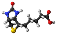
| + | '''Streptavidin''', from the bacteria ''Streptomyces avidinii'', is a tetrameric protein that has a strong affinity for binding '''biotin''' (the 3D structure of <scene name='58/580641/Biotin_bs/1'>biotin</scene> is now displayed at the right-hand panel). | ||
| + | {| | ||
| + | |[[Image:Biotin structure.png]] | ||
| + | |align="center"| Biotin | ||
| + | |[[Image:Biotin 3D balls.png]] | ||
| + | |} | ||
| - | + | You can read more details at the [[Streptavidin|avidin and streptavidin page]] | |
| - | You can read more details at [[Streptavidin| | + | |
This page focuses on the binding site for biotin and how two of the subunits collaborate in binding. | This page focuses on the binding site for biotin and how two of the subunits collaborate in binding. | ||
| - | The <scene name=' | + | The <scene name='58/580641/One_subunit/1'>first display</scene> shows a single subunit of streptavidin with its bound biotin. |
| - | What does the binding site look like? Let's display the <scene name=' | + | What does the binding site look like? Let's display the <scene name='58/580641/One_subunit_solid/1'>solid volume of the protein</scene> (in pale blue). |
As you see, biotin is embedded inside streptavidin, but a considerable –and hydrophobic– part of the biotin molecule remains exposed. | As you see, biotin is embedded inside streptavidin, but a considerable –and hydrophobic– part of the biotin molecule remains exposed. | ||
| - | However, when we add the <scene name=' | + | However, when we add the <scene name='58/580641/One_subunit_solid_solidcover/1'>portion of the neighbouring streptavidin subunit</scene> (residues 116 to 121) that contacts with the first subunit, you will see that it covers most of the biotin molecule. |
Now only the carboxylate group of biotin (note the two red oxygens) sticks out. | Now only the carboxylate group of biotin (note the two red oxygens) sticks out. | ||
Current revision
Streptavidin binds biotin with extraordinary affinity
| |||||||||||


