We apologize for Proteopedia being slow to respond. For the past two years, a new implementation of Proteopedia has been being built. Soon, it will replace this 18-year old system. All existing content will be moved to the new system at a date that will be announced here.
4p3r
From Proteopedia
(Difference between revisions)
| (9 intermediate revisions not shown.) | |||
| Line 1: | Line 1: | ||
| - | '''Unreleased structure''' | ||
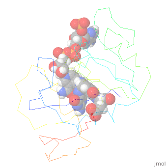
| - | + | ==Cryogenic WT DHFR, time-averaged ensemble== | |
| + | <StructureSection load='4p3r' size='340' side='right'caption='[[4p3r]], [[Resolution|resolution]] 1.15Å' scene=''> | ||
| + | == Structural highlights == | ||
| + | <table><tr><td colspan='2'>[[4p3r]] is a 1 chain structure with sequence from [https://en.wikipedia.org/wiki/Escherichia_coli_str._K-12_substr._DH10B Escherichia coli str. K-12 substr. DH10B]. Full crystallographic information is available from [http://oca.weizmann.ac.il/oca-bin/ocashort?id=4P3R OCA]. For a <b>guided tour on the structure components</b> use [https://proteopedia.org/fgij/fg.htm?mol=4P3R FirstGlance]. <br> | ||
| + | </td></tr><tr id='method'><td class="sblockLbl"><b>[[Empirical_models|Method:]]</b></td><td class="sblockDat" id="methodDat">X-ray diffraction, [[Resolution|Resolution]] 1.15Å</td></tr> | ||
| + | <tr id='ligand'><td class="sblockLbl"><b>[[Ligand|Ligands:]]</b></td><td class="sblockDat" id="ligandDat"><scene name='pdbligand=FOL:FOLIC+ACID'>FOL</scene>, <scene name='pdbligand=NAP:NADP+NICOTINAMIDE-ADENINE-DINUCLEOTIDE+PHOSPHATE'>NAP</scene></td></tr> | ||
| + | <tr id='resources'><td class="sblockLbl"><b>Resources:</b></td><td class="sblockDat"><span class='plainlinks'>[https://proteopedia.org/fgij/fg.htm?mol=4p3r FirstGlance], [http://oca.weizmann.ac.il/oca-bin/ocaids?id=4p3r OCA], [https://pdbe.org/4p3r PDBe], [https://www.rcsb.org/pdb/explore.do?structureId=4p3r RCSB], [https://www.ebi.ac.uk/pdbsum/4p3r PDBsum], [https://prosat.h-its.org/prosat/prosatexe?pdbcode=4p3r ProSAT]</span></td></tr> | ||
| + | </table> | ||
| + | <div style="background-color:#fffaf0;"> | ||
| + | == Publication Abstract from PubMed == | ||
| + | Most macromolecular X-ray structures are determined from cryocooled crystals, but it is unclear whether cryocooling distorts functionally relevant flexibility. Here we compare independently acquired pairs of high-resolution data sets of a model Michaelis complex of dihydrofolate reductase (DHFR), collected by separate groups at both room and cryogenic temperatures. These data sets allow us to isolate the differences between experimental procedures and between temperatures. Our analyses of multiconformer models and time-averaged ensembles suggest that cryocooling suppresses and otherwise modifies side-chain and main-chain conformational heterogeneity, quenching dynamic contact networks. Despite some idiosyncratic differences, most changes from room temperature to cryogenic temperature are conserved and likely reflect temperature-dependent solvent remodeling. Both cryogenic data sets point to additional conformations not evident in the corresponding room temperature data sets, suggesting that cryocooling does not merely trap preexisting conformational heterogeneity. Our results demonstrate that crystal cryocooling consistently distorts the energy landscape of DHFR, a paragon for understanding functional protein dynamics. | ||
| - | + | Crystal Cryocooling Distorts Conformational Heterogeneity in a Model Michaelis Complex of DHFR.,Keedy DA, van den Bedem H, Sivak DA, Petsko GA, Ringe D, Wilson MA, Fraser JS Structure. 2014 Jun 10;22(6):899-910. doi: 10.1016/j.str.2014.04.016. Epub 2014, May 29. PMID:24882744<ref>PMID:24882744</ref> | |
| - | + | From MEDLINE®/PubMed®, a database of the U.S. National Library of Medicine.<br> | |
| + | </div> | ||
| + | <div class="pdbe-citations 4p3r" style="background-color:#fffaf0;"></div> | ||
| + | |||
| + | ==See Also== | ||
| + | *[[Dihydrofolate reductase 3D structures|Dihydrofolate reductase 3D structures]] | ||
| + | == References == | ||
| + | <references/> | ||
| + | __TOC__ | ||
| + | </StructureSection> | ||
| + | [[Category: Escherichia coli str. K-12 substr. DH10B]] | ||
| + | [[Category: Large Structures]] | ||
| + | [[Category: Fraser JS]] | ||
| + | [[Category: Keedy DA]] | ||
| + | [[Category: Van den Bedem H]] | ||
Current revision
Cryogenic WT DHFR, time-averaged ensemble
| |||||||||||