This old version of Proteopedia is provided for student assignments while the new version is undergoing repairs. Content and edits done in this old version of Proteopedia after March 1, 2026 will eventually be lost when it is retired in about June of 2026.
Apply for new accounts at the new Proteopedia. Your logins will work in both the old and new versions.
4op0
From Proteopedia
(Difference between revisions)
| (5 intermediate revisions not shown.) | |||
| Line 1: | Line 1: | ||
| + | |||
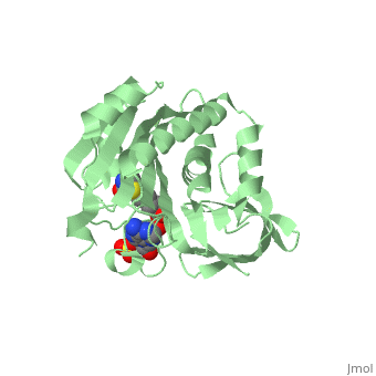
==Crystal structure of biotin protein ligase (RV3279C) of Mycobacterium tuberculosis, complexed with biotinyl-5'-AMP== | ==Crystal structure of biotin protein ligase (RV3279C) of Mycobacterium tuberculosis, complexed with biotinyl-5'-AMP== | ||
| - | <StructureSection load='4op0' size='340' side='right' caption='[[4op0]], [[Resolution|resolution]] 1.70Å' scene=''> | + | <StructureSection load='4op0' size='340' side='right'caption='[[4op0]], [[Resolution|resolution]] 1.70Å' scene=''> |
== Structural highlights == | == Structural highlights == | ||
| - | <table><tr><td colspan='2'>[[4op0]] is a 2 chain structure. Full crystallographic information is available from [http://oca.weizmann.ac.il/oca-bin/ocashort?id=4OP0 OCA]. <br> | + | <table><tr><td colspan='2'>[[4op0]] is a 2 chain structure with sequence from [https://en.wikipedia.org/wiki/Mycobacterium_tuberculosis_H37Rv Mycobacterium tuberculosis H37Rv]. Full crystallographic information is available from [http://oca.weizmann.ac.il/oca-bin/ocashort?id=4OP0 OCA]. For a <b>guided tour on the structure components</b> use [https://proteopedia.org/fgij/fg.htm?mol=4OP0 FirstGlance]. <br> |
| - | </td></tr><tr><td class="sblockLbl"><b>[[ | + | </td></tr><tr id='method'><td class="sblockLbl"><b>[[Empirical_models|Method:]]</b></td><td class="sblockDat" id="methodDat">X-ray diffraction, [[Resolution|Resolution]] 1.7Å</td></tr> |
| - | <tr><td class="sblockLbl"><b> | + | <tr id='ligand'><td class="sblockLbl"><b>[[Ligand|Ligands:]]</b></td><td class="sblockDat" id="ligandDat"><scene name='pdbligand=BT5:BIOTINYL-5-AMP'>BT5</scene>, <scene name='pdbligand=SO4:SULFATE+ION'>SO4</scene></td></tr> |
| - | <tr><td class="sblockLbl"><b>Resources:</b></td><td class="sblockDat"><span class='plainlinks'>[ | + | <tr id='resources'><td class="sblockLbl"><b>Resources:</b></td><td class="sblockDat"><span class='plainlinks'>[https://proteopedia.org/fgij/fg.htm?mol=4op0 FirstGlance], [http://oca.weizmann.ac.il/oca-bin/ocaids?id=4op0 OCA], [https://pdbe.org/4op0 PDBe], [https://www.rcsb.org/pdb/explore.do?structureId=4op0 RCSB], [https://www.ebi.ac.uk/pdbsum/4op0 PDBsum], [https://prosat.h-its.org/prosat/prosatexe?pdbcode=4op0 ProSAT]</span></td></tr> |
| - | <table> | + | </table> |
| + | == Function == | ||
| + | [https://www.uniprot.org/uniprot/BIRA_MYCTU BIRA_MYCTU] Catalyzes the transfer of biotin onto a conserved lysine residue of the biotin carboxyl carrier protein (BCCP) domain of acetyl-CoA carboxylase and converts it to active holo-BCCP (PubMed:18509457, PubMed:24723382). Forms an acyl-adenylate intermediate (PubMed:18509457, PubMed:24723382). Cannot use GTP or desthiobiotin (PubMed:18509457).<ref>PMID:18509457</ref> <ref>PMID:24723382</ref> | ||
<div style="background-color:#fffaf0;"> | <div style="background-color:#fffaf0;"> | ||
== Publication Abstract from PubMed == | == Publication Abstract from PubMed == | ||
| Line 13: | Line 16: | ||
Active site conformational changes upon reaction intermediate biotinyl-5'-AMP binding in biotin protein ligase from Mycobacterium tuberculosis.,Ma Q, Akhter Y, Wilmanns M, Ehebauer MT Protein Sci. 2014 Apr 9. doi: 10.1002/pro.2475. PMID:24723382<ref>PMID:24723382</ref> | Active site conformational changes upon reaction intermediate biotinyl-5'-AMP binding in biotin protein ligase from Mycobacterium tuberculosis.,Ma Q, Akhter Y, Wilmanns M, Ehebauer MT Protein Sci. 2014 Apr 9. doi: 10.1002/pro.2475. PMID:24723382<ref>PMID:24723382</ref> | ||
| - | From | + | From MEDLINE®/PubMed®, a database of the U.S. National Library of Medicine.<br> |
</div> | </div> | ||
| + | <div class="pdbe-citations 4op0" style="background-color:#fffaf0;"></div> | ||
| + | |||
| + | ==See Also== | ||
| + | *[[Biotin Protein Ligase|Biotin Protein Ligase]] | ||
| + | *[[Biotin Protein Ligase 3D structures|Biotin Protein Ligase 3D structures]] | ||
== References == | == References == | ||
<references/> | <references/> | ||
__TOC__ | __TOC__ | ||
</StructureSection> | </StructureSection> | ||
| - | [[Category: | + | [[Category: Large Structures]] |
| - | [[Category: | + | [[Category: Mycobacterium tuberculosis H37Rv]] |
| - | [[Category: | + | [[Category: Akhter Y]] |
| - | [[Category: | + | [[Category: Ma Q]] |
| - | [[Category: | + | [[Category: Wilmanns M]] |
Current revision
Crystal structure of biotin protein ligase (RV3279C) of Mycobacterium tuberculosis, complexed with biotinyl-5'-AMP
| |||||||||||