This old version of Proteopedia is provided for student assignments while the new version is undergoing repairs. Content and edits done in this old version of Proteopedia after March 1, 2026 will eventually be lost when it is retired in about June of 2026.
Apply for new accounts at the new Proteopedia. Your logins will work in both the old and new versions.
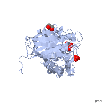
3vnz
From Proteopedia
(Difference between revisions)
| (4 intermediate revisions not shown.) | |||
| Line 1: | Line 1: | ||
| + | |||
==Crystal structure of beta-glucuronidase from Acidobacterium capsulatum in complex with D-glucuronic acid== | ==Crystal structure of beta-glucuronidase from Acidobacterium capsulatum in complex with D-glucuronic acid== | ||
| - | <StructureSection load='3vnz' size='340' side='right' caption='[[3vnz]], [[Resolution|resolution]] 1.80Å' scene=''> | + | <StructureSection load='3vnz' size='340' side='right'caption='[[3vnz]], [[Resolution|resolution]] 1.80Å' scene=''> |
== Structural highlights == | == Structural highlights == | ||
| - | <table><tr><td colspan='2'>[[3vnz]] is a 1 chain structure with sequence from [ | + | <table><tr><td colspan='2'>[[3vnz]] is a 1 chain structure with sequence from [https://en.wikipedia.org/wiki/Acidobacterium_capsulatum_ATCC_51196 Acidobacterium capsulatum ATCC 51196]. Full crystallographic information is available from [http://oca.weizmann.ac.il/oca-bin/ocashort?id=3VNZ OCA]. For a <b>guided tour on the structure components</b> use [https://proteopedia.org/fgij/fg.htm?mol=3VNZ FirstGlance]. <br> |
| - | </td></tr><tr><td class="sblockLbl"><b>[[ | + | </td></tr><tr id='method'><td class="sblockLbl"><b>[[Empirical_models|Method:]]</b></td><td class="sblockDat" id="methodDat">X-ray diffraction, [[Resolution|Resolution]] 1.8Å</td></tr> |
| - | + | <tr id='ligand'><td class="sblockLbl"><b>[[Ligand|Ligands:]]</b></td><td class="sblockDat" id="ligandDat"><scene name='pdbligand=BDP:BETA-D-GLUCOPYRANURONIC+ACID'>BDP</scene>, <scene name='pdbligand=GOL:GLYCEROL'>GOL</scene>, <scene name='pdbligand=PO4:PHOSPHATE+ION'>PO4</scene></td></tr> | |
| - | <tr><td class="sblockLbl"><b>[[ | + | <tr id='resources'><td class="sblockLbl"><b>Resources:</b></td><td class="sblockDat"><span class='plainlinks'>[https://proteopedia.org/fgij/fg.htm?mol=3vnz FirstGlance], [http://oca.weizmann.ac.il/oca-bin/ocaids?id=3vnz OCA], [https://pdbe.org/3vnz PDBe], [https://www.rcsb.org/pdb/explore.do?structureId=3vnz RCSB], [https://www.ebi.ac.uk/pdbsum/3vnz PDBsum], [https://prosat.h-its.org/prosat/prosatexe?pdbcode=3vnz ProSAT]</span></td></tr> |
| - | + | </table> | |
| - | <tr><td class="sblockLbl"><b>Resources:</b></td><td class="sblockDat"><span class='plainlinks'>[ | + | == Function == |
| - | <table> | + | [https://www.uniprot.org/uniprot/C1F2K5_ACIC5 C1F2K5_ACIC5] |
| + | <div style="background-color:#fffaf0;"> | ||
| + | == Publication Abstract from PubMed == | ||
| + | We present the first structure of a glycoside hydrolase family 79 beta-glucuronidase from Acidobacterium capsulatum, both as a product complex with beta-D-glucuronic acid (GlcA) and as its trapped covalent 2-fluoroglucuronyl intermediate. This enzyme consists of a catalytic (beta/alpha)(8)-barrel domain and a beta-domain with irregular Greek key motifs that is of unknown function. The enzyme showed beta-glucuronidase activity and trace levels of beta-glucosidase and beta-xylosidase activities. In conjunction with mutagenesis studies, these structures identify the catalytic residues as Glu(173) (acid base) and Glu(287) (nucleophile), consistent with the retaining mechanism demonstrated by (1)H NMR analysis. Glu(45), Tyr(243), Tyr(292)-Gly(294), and Tyr(334) form the catalytic pocket and provide substrate discrimination. Consistent with this, the Y292A mutation, which affects the interaction between the main chains of Gln(293) and Gly(294) and the GlcA carboxyl group, resulted in significant loss of beta-glucuronidase activity while retaining the side activities at wild-type levels. Likewise, although the beta-glucuronidase activity of the Y334F mutant is ~200-fold lower (k(cat)/K(m)) than that of the wild-type enzyme, the beta-glucosidase activity is actually 3 times higher and the beta-xylosidase activity is only 2.5-fold lower than the equivalent parameters for wild type, consistent with a role for Tyr(334) in recognition of the C6 position of GlcA. The involvement of Glu(45) in discriminating against binding of the O-methyl group at the C4 position of GlcA is revealed in the fact that the E45D mutant hydrolyzes PNP-beta-GlcA approximately 300-fold slower (k(cat)/K(m)) than does the wild-type enzyme, whereas 4-O-methyl-GlcA-containing oligosaccharides are hydrolyzed only 7-fold slower. | ||
| + | |||
| + | Structural and biochemical characterization of glycoside hydrolase family 79 beta-glucuronidase from Acidobacterium capsulatum.,Michikawa M, Ichinose H, Momma M, Biely P, Jongkees S, Yoshida M, Kotake T, Tsumuraya Y, Withers SG, Fujimoto Z, Kaneko S J Biol Chem. 2012 Apr 20;287(17):14069-77. Epub 2012 Feb 24. PMID:22367201<ref>PMID:22367201</ref> | ||
| + | |||
| + | From MEDLINE®/PubMed®, a database of the U.S. National Library of Medicine.<br> | ||
| + | </div> | ||
| + | <div class="pdbe-citations 3vnz" style="background-color:#fffaf0;"></div> | ||
| + | |||
| + | ==See Also== | ||
| + | *[[Glucuronidase|Glucuronidase]] | ||
| + | *[[Glucuronisidase 3D structures|Glucuronisidase 3D structures]] | ||
| + | == References == | ||
| + | <references/> | ||
__TOC__ | __TOC__ | ||
</StructureSection> | </StructureSection> | ||
| - | [[Category: Acidobacterium capsulatum]] | + | [[Category: Acidobacterium capsulatum ATCC 51196]] |
| - | [[Category: | + | [[Category: Large Structures]] |
| - | [[Category: Biely | + | [[Category: Biely P]] |
| - | [[Category: Fujimoto | + | [[Category: Fujimoto Z]] |
| - | [[Category: Ichinose | + | [[Category: Ichinose H]] |
| - | [[Category: Kaneko | + | [[Category: Kaneko S]] |
| - | [[Category: Kotake | + | [[Category: Kotake Y]] |
| - | [[Category: Michikawa | + | [[Category: Michikawa M]] |
| - | [[Category: Momma | + | [[Category: Momma M]] |
| - | [[Category: Tsumuraya | + | [[Category: Tsumuraya Y]] |
| - | [[Category: Yoshida | + | [[Category: Yoshida M]] |
| - | + | ||
| - | + | ||
| - | + | ||
| - | + | ||
Current revision
Crystal structure of beta-glucuronidase from Acidobacterium capsulatum in complex with D-glucuronic acid
| |||||||||||