Hila Cohen/Test Page
From Proteopedia
(Difference between revisions)
| (37 intermediate revisions not shown.) | |||
| Line 1: | Line 1: | ||
| - | ==Avidin (Structure)== | ||
| - | <StructureSection load='1VYO' size='250' side='left' caption='CRYSTAL STRUCTURE OF AVIDIN'=> | ||
| - | This is a default text for your page '''Hila Cohen/Test Page'''. Click above on '''edit this page''' to modify. Be careful with the < and > signs. | ||
| - | You may include any references to papers as in: the use of JSmol in Proteopedia <ref>DOI 10.1002/ijch.201300024</ref> or to the article describing Jmol <ref>PMID:21638687</ref> to the rescue. | ||
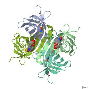
| - | == | + | <StructureSection load='1AVD' size='300' side='left' caption='Crystal Structure of Avidin With Biotin (PDB code [[1avd]])'> |
| - | = | + | '''Avidin''' is a protein that’s bind Vitamin B7, <scene name='60/607867/Biotin/2'>Biotin</scene>. |
| + | The Avidin is produced in the ovary of some lay eggs animals, and it’s found in the white of the egg <ref>doi:10.1016/S0065-3233(08)60411-8</ref>. | ||
| - | == | + | == Structure == |
| - | + | Avidin is a tetrameric protein, but here will be shown only two units of the whole structure just to simplify it. | |
| - | + | The secondary structure of each Avidin monomer combines <scene name='60/607867/A-helix/1'>alpha-helix</scene> (pink) and an 8 stranded antiparalle <scene name='60/607867/Beta-helix/1'>beta-sheets</scene> (turquoise). The tertiary structure of each monomer is beta-barrel <ref>PMID: 8506353</ref>. | |
| + | |||
| + | == Structural highlights == | ||
| + | Because of the curved structure of the protein, it’s easier to see where the N terminus is begins and the C terminus ends with <scene name='60/607867/N_to_c_rainbow/1'>Rainbow color display</scene>. | ||
| + | {{Template:ColorKey_Amino2CarboxyRainbow}} | ||
| - | </StructureSection> | ||
== References == | == References == | ||
<references/> | <references/> | ||
Current revision
| |||||||||||