We apologize for Proteopedia being slow to respond. For the past two years, a new implementation of Proteopedia has been being built. Soon, it will replace this 18-year old system. All existing content will be moved to the new system at a date that will be announced here.
Chameleon peptide
From Proteopedia
(Difference between revisions)
| (One intermediate revision not shown.) | |||
| Line 1: | Line 1: | ||
==Does the amino acid sequence ''really'' determine the 3D structure of a peptide?== | ==Does the amino acid sequence ''really'' determine the 3D structure of a peptide?== | ||
| - | <StructureSection load='2gb1' size='350' side='right' caption='2gb1' scene='61/611445/Rainbow_cartoon/1'> | + | <StructureSection load='2gb1' size='350' side='right' caption='Protein G immunoglobulin binding domain (PDB code [[2gb1]])' scene='61/611445/Rainbow_cartoon/1'> |
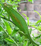
[[Image:Chameleon.jpg|left|170px|thumb|Chamelion]] | [[Image:Chameleon.jpg|left|170px|thumb|Chamelion]] | ||
==General question== | ==General question== | ||
| Line 9: | Line 9: | ||
== Seeing is believing == | == Seeing is believing == | ||
| - | To simplify the figure, the entire IgG-Binding domain<ref>PMID:1871600 </ref> is colored <scene name='61/611445/Cartoon_beige/1'>beige</scene>. Now, if the amino acids residues, <scene name='61/611445/ | + | To simplify the figure, the entire IgG-Binding domain<ref>PMID:1871600 </ref> is colored <scene name='61/611445/Cartoon_beige/1'>beige</scene>. Now, if the amino acids residues, <<scene name='61/611445/Cartoon_blanche/2'>23-33</scene>>, are changed to the ''Chameleon'' sequence i.e. <span style="color:#FFFFFF; background:#FF69B4">AWTVEKAFKTF</span style>, virtually no change in structure is seen. |
Specifically the WT sequence in yellow (residues 23-33) is changed to Chameleon sequence shown in pink, , n.b. only 5 AA's have been changed in this region<br /> | Specifically the WT sequence in yellow (residues 23-33) is changed to Chameleon sequence shown in pink, , n.b. only 5 AA's have been changed in this region<br /> | ||
Current revision
Does the amino acid sequence really determine the 3D structure of a peptide?
| |||||||||||
References
- ↑ Minor DL Jr, Kim PS. Context-dependent secondary structure formation of a designed protein sequence. Nature. 1996 Apr 25;380(6576):730-4. PMID:8614471 doi:http://dx.doi.org/10.1038/380730a0
- ↑ Zhou X, Alber F, Folkers G, Gonnet GH, Chelvanayagam G. An analysis of the helix-to-strand transition between peptides with identical sequence. Proteins. 2000 Nov 1;41(2):248-56. PMID:10966577
- ↑ Gronenborn AM, Filpula DR, Essig NZ, Achari A, Whitlow M, Wingfield PT, Clore GM. A novel, highly stable fold of the immunoglobulin binding domain of streptococcal protein G. Science. 1991 Aug 9;253(5020):657-61. PMID:1871600

