We apologize for Proteopedia being slow to respond. For the past two years, a new implementation of Proteopedia has been being built. Soon, it will replace this 18-year old system. All existing content will be moved to the new system at a date that will be announced here.
Sandbox Reserved 960
From Proteopedia
(Difference between revisions)
| (7 intermediate revisions not shown.) | |||
| Line 5: | Line 5: | ||
<nowiki> | <nowiki> | ||
| - | The protein AmelASP1 has been identified in the antennae from the honeybee ''Apis mellifera''. Its primary sequence is a 144 amino acids polypeptide with a molecular weight of 13.180 kDa. AmelASP1 is part of the Pheromone Binding Protein (PBP) family. The 3D representation shown below was obtained at pH 5.5 using the [http://www-dsv.cea.fr/en/life-science-div/all-the-news/scientific-results/nanodrops-for-bioactive-compound-synthesis-and-screening nano-drops technique]. | + | The protein '''AmelASP1''' has been identified in the antennae from the honeybee ''Apis mellifera''. Its primary sequence is a 144 amino acids polypeptide with a molecular weight of 13.180 kDa. AmelASP1 is part of the '''Pheromone Binding Protein (PBP)''' family. The 3D representation shown below was obtained at pH 5.5 using the [http://www-dsv.cea.fr/en/life-science-div/all-the-news/scientific-results/nanodrops-for-bioactive-compound-synthesis-and-screening nano-drops technique]. |
| Line 51: | Line 51: | ||
=== pH influence === | === pH influence === | ||
| - | pH affects the flexibility of ASP1 because it induces a different protonation state of the <scene name='60/604479/Ionizable_residues/1'>ionizable residues</scene>. Protonated residues | + | pH affects the flexibility of ASP1 because it induces a different protonation state of the <scene name='60/604479/Ionizable_residues/1'>ionizable residues</scene>. Protonated residues give rise to micro-environnment changes which propagate all along the protein. Consequently, ASP1 is no longer able to interact with its ligands even if ionizable residues are distant from the cavity. <ref>PMID: 19481550</ref> |
In fact, depending of the pH level, Asp35 bend the C terminal domain against the cavity. | In fact, depending of the pH level, Asp35 bend the C terminal domain against the cavity. | ||
At pH 5.5, <scene name='60/604479/C-term_asp35/1'>Asp 35</scene> is protonated and C terminal domain isn’t bend against the cavity. While ASP1 is a monomere at acid pH, it can dimerize at neutral and basic pH.<ref>PMID: 25337796</ref> | At pH 5.5, <scene name='60/604479/C-term_asp35/1'>Asp 35</scene> is protonated and C terminal domain isn’t bend against the cavity. While ASP1 is a monomere at acid pH, it can dimerize at neutral and basic pH.<ref>PMID: 25337796</ref> | ||
| Line 57: | Line 57: | ||
== Ligands == | == Ligands == | ||
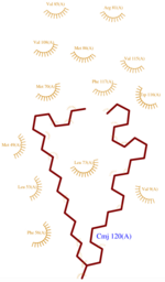
| - | [[Image:CMJ_Ligplot.png|150px|right|thumb|'''Fig.1''' CMJ Ligplot]] | + | [[Image:CMJ_Ligplot.png|150px|right|thumb|'''Fig.1''' CMJ Ligplot<ref>http://www.ebi.ac.uk/thornton-srv/databases/cgi-bin/pdbsum/GetPage.pl?pdbcode=3fe6&template=ligands.html&l=1.1</ref>]] |
In order to determine this {{Template:ColorKey Composition Protein}}’s structure, several {{Template:ColorKey Composition Ligand}} has been used at pH 5.5 because this low pH fits with its natural medium in the bee antenna. | In order to determine this {{Template:ColorKey Composition Protein}}’s structure, several {{Template:ColorKey Composition Ligand}} has been used at pH 5.5 because this low pH fits with its natural medium in the bee antenna. | ||
The three ligands used to characterize and purify AmelASP1 are : | The three ligands used to characterize and purify AmelASP1 are : | ||
| - | *<scene name='60/604479/Cmj/3'>CMJ</scene> also known as (20s)-20-Methyldotetracontane, is a serendipitous ligand. This term signify that the purification of this molecule was completely fortuitous. It is a big unsaturated mono-methyl branched carbone chain with formula C43H88. This ligand fits in the {{Template:ColorKey_Hydrophobic}} cavity of AmelASP1 thanks to several interactions with <scene name='60/604479/Cmj_binding_residues/ | + | *<scene name='60/604479/Cmj/3'>CMJ</scene> also known as (20s)-20-Methyldotetracontane, is a serendipitous ligand. This term signify that the purification of this molecule was completely fortuitous. It is a big unsaturated mono-methyl branched carbone chain with formula C43H88. This ligand fits in the {{Template:ColorKey_Hydrophobic}} cavity of AmelASP1 thanks to several interactions with <scene name='60/604479/Cmj_binding_residues/6'>specific residues.</scene> ('''Fig.1''') |
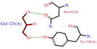
| - | *[[Image:GOL_Ligplot.png|200px|left|thumb|'''Fig.2''' GOL Ligplot]]<scene name='60/604479/Gol/1'>Glycerol</scene> (C3H8O3) also known as GOL, is a ligand used for cryoprotection during the purification process of the protein. It is supposedly helping the main ligand to reach its binding site. | + | *[[Image:GOL_Ligplot.png|200px|left|thumb|'''Fig.2''' GOL Ligplot<ref>http://www.ebi.ac.uk/thornton-srv/databases/cgi-bin/pdbsum/GetPage.pl?pdbcode=3fe6&template=ligands.html&l=2.1</ref>]]<scene name='60/604479/Gol/1'>Glycerol</scene> (C3H8O3) also known as GOL, is a ligand used for cryoprotection during the purification process of the protein. It is supposedly helping the main ligand to reach its binding site. |
| - | To do so, GOL links to<scene name='60/604479/Gol_binding_residues/1'> Asn 41 and Tyr 102.</scene> | + | To do so, GOL links to<scene name='60/604479/Gol_binding_residues/1'> Asn 41 and Tyr 102.</scene> ('''Fig.2''') |
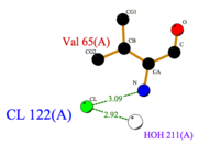
| - | [[Image:Cl_Ligplot.png|right|thumb|'''Fig.3''' Cl Ligplot]] | + | [[Image:Cl_Ligplot.png|right|thumb|'''Fig.3''' Cl Ligplot<ref>http://www.ebi.ac.uk/thornton-srv/databases/cgi-bin/pdbsum/GetPage.pl?pdbcode=3fe6&template=ligands.html&o=METAL&l=1.1</ref>]] |
| - | *<scene name='60/604479/Cl/1'>Chloride ion</scene> facilitates the binding of other ligands to the protein. Its abundance around ASP1 varies according to changing pH conditions. At pH 5.5, <scene name='60/604479/Cl_binding_residue/1'>Val 65</scene> is the only amino acid able to fix a chloride ion. | + | *<scene name='60/604479/Cl/1'>Chloride ion</scene> facilitates the binding of other ligands to the protein. Its abundance around ASP1 varies according to changing pH conditions. At pH 5.5, <scene name='60/604479/Cl_binding_residue/1'>Val 65</scene> is here the only amino acid able to fix a chloride ion. ('''Fig.3''') |
However, in natural conditions, binding ligands are the pheromones secreted by the queen such as 9-ODA. | However, in natural conditions, binding ligands are the pheromones secreted by the queen such as 9-ODA. | ||
| Line 99: | Line 99: | ||
== Contributors == | == Contributors == | ||
| - | + | ||
Sophie Morin & Mathias Buytaert | Sophie Morin & Mathias Buytaert | ||
Current revision
| This Sandbox is Reserved from 15/11/2014, through 15/05/2015 for use in the course "Biomolecule" taught by Bruno Kieffer at the Strasbourg University. This reservation includes Sandbox Reserved 951 through Sandbox Reserved 975. |
To get started:
More help: Help:Editing |
Antennal Specific Protein-1 from Apis mellifera (AmelASP1) with a serendipitous ligand at pH 5.5
| |||||||||||
Contributors
Sophie Morin & Mathias Buytaert
References for further information on the pheromone binding protein from Apis mellifera
- ↑ http://www.genome.jp/dbget-bin/www_bget?pdb:3FE6
- ↑ Pesenti ME, Spinelli S, Bezirard V, Briand L, Pernollet JC, Tegoni M, Cambillau C. Structural basis of the honey bee PBP pheromone and pH-induced conformational change. J Mol Biol. 2008 Jun 27;380(1):158-69. Epub 2008 Apr 27. PMID:18508083 doi:10.1016/j.jmb.2008.04.048
- ↑ Han L, Zhang YJ, Zhang L, Cui X, Yu J, Zhang Z, Liu MS. Operating mechanism and molecular dynamics of pheromone-binding protein ASP1 as influenced by pH. PLoS One. 2014 Oct 22;9(10):e110565. doi: 10.1371/journal.pone.0110565., eCollection 2014. PMID:25337796 doi:http://dx.doi.org/10.1371/journal.pone.0110565
- ↑ Lartigue A, Gruez A, Briand L, Blon F, Bezirard V, Walsh M, Pernollet JC, Tegoni M, Cambillau C. Sulfur single-wavelength anomalous diffraction crystal structure of a pheromone-binding protein from the honeybee Apis mellifera L. J Biol Chem. 2004 Feb 6;279(6):4459-64. Epub 2003 Oct 31. PMID:14594955 doi:10.1074/jbc.M311212200
- ↑ Pesenti ME, Spinelli S, Bezirard V, Briand L, Pernollet JC, Campanacci V, Tegoni M, Cambillau C. Queen bee pheromone binding protein pH-induced domain swapping favors pheromone release. J Mol Biol. 2009 Jul 31;390(5):981-90. Epub 2009 May 28. PMID:19481550 doi:10.1016/j.jmb.2009.05.067
- ↑ Han L, Zhang YJ, Zhang L, Cui X, Yu J, Zhang Z, Liu MS. Operating mechanism and molecular dynamics of pheromone-binding protein ASP1 as influenced by pH. PLoS One. 2014 Oct 22;9(10):e110565. doi: 10.1371/journal.pone.0110565., eCollection 2014. PMID:25337796 doi:http://dx.doi.org/10.1371/journal.pone.0110565
- ↑ http://www.ebi.ac.uk/thornton-srv/databases/cgi-bin/pdbsum/GetPage.pl?pdbcode=3fe6&template=ligands.html&l=1.1
- ↑ http://www.ebi.ac.uk/thornton-srv/databases/cgi-bin/pdbsum/GetPage.pl?pdbcode=3fe6&template=ligands.html&l=2.1
- ↑ http://www.ebi.ac.uk/thornton-srv/databases/cgi-bin/pdbsum/GetPage.pl?pdbcode=3fe6&template=ligands.html&o=METAL&l=1.1


