We apologize for Proteopedia being slow to respond. For the past two years, a new implementation of Proteopedia has been being built. Soon, it will replace this 18-year old system. All existing content will be moved to the new system at a date that will be announced here.
2h24
From Proteopedia
(Difference between revisions)
| (15 intermediate revisions not shown.) | |||
| Line 1: | Line 1: | ||
| - | [[Image:2h24.gif|left|200px]] | ||
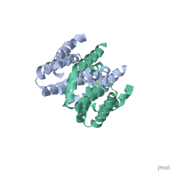
| - | + | ==Crystal structure of human IL-10== | |
| - | + | <StructureSection load='2h24' size='340' side='right'caption='[[2h24]], [[Resolution|resolution]] 2.00Å' scene=''> | |
| - | + | == Structural highlights == | |
| - | + | <table><tr><td colspan='2'>[[2h24]] is a 1 chain structure with sequence from [https://en.wikipedia.org/wiki/Homo_sapiens Homo sapiens]. Full crystallographic information is available from [http://oca.weizmann.ac.il/oca-bin/ocashort?id=2H24 OCA]. For a <b>guided tour on the structure components</b> use [https://proteopedia.org/fgij/fg.htm?mol=2H24 FirstGlance]. <br> | |
| - | + | </td></tr><tr id='method'><td class="sblockLbl"><b>[[Empirical_models|Method:]]</b></td><td class="sblockDat" id="methodDat">X-ray diffraction, [[Resolution|Resolution]] 2Å</td></tr> | |
| - | | | + | <tr id='resources'><td class="sblockLbl"><b>Resources:</b></td><td class="sblockDat"><span class='plainlinks'>[https://proteopedia.org/fgij/fg.htm?mol=2h24 FirstGlance], [http://oca.weizmann.ac.il/oca-bin/ocaids?id=2h24 OCA], [https://pdbe.org/2h24 PDBe], [https://www.rcsb.org/pdb/explore.do?structureId=2h24 RCSB], [https://www.ebi.ac.uk/pdbsum/2h24 PDBsum], [https://prosat.h-its.org/prosat/prosatexe?pdbcode=2h24 ProSAT]</span></td></tr> |
| - | + | </table> | |
| - | + | == Function == | |
| - | + | [https://www.uniprot.org/uniprot/IL10_HUMAN IL10_HUMAN] Inhibits the synthesis of a number of cytokines, including IFN-gamma, IL-2, IL-3, TNF and GM-CSF produced by activated macrophages and by helper T-cells. | |
| - | + | == Evolutionary Conservation == | |
| - | + | [[Image:Consurf_key_small.gif|200px|right]] | |
| - | == | + | Check<jmol> |
| + | <jmolCheckbox> | ||
| + | <scriptWhenChecked>; select protein; define ~consurf_to_do selected; consurf_initial_scene = true; script "/wiki/ConSurf/h2/2h24_consurf.spt"</scriptWhenChecked> | ||
| + | <scriptWhenUnchecked>script /wiki/extensions/Proteopedia/spt/initialview03.spt</scriptWhenUnchecked> | ||
| + | <text>to colour the structure by Evolutionary Conservation</text> | ||
| + | </jmolCheckbox> | ||
| + | </jmol>, as determined by [http://consurfdb.tau.ac.il/ ConSurfDB]. You may read the [[Conservation%2C_Evolutionary|explanation]] of the method and the full data available from [http://bental.tau.ac.il/new_ConSurfDB/main_output.php?pdb_ID=2h24 ConSurf]. | ||
| + | <div style="clear:both"></div> | ||
| + | <div style="background-color:#fffaf0;"> | ||
| + | == Publication Abstract from PubMed == | ||
Interleukin-10 receptor 2 (IL-10R2) is a critical component of the IL-10.IL-10R1.IL-10R2 complex which regulates IL-10-mediated immunomodulatory responses. The ternary IL-10 signaling complex is assembled in a sequential order with the IL-10.IL-10R1 interaction occurring first followed by engagement of the IL-10R2 chain. In this study we map the IL-10R2 binding site on IL-10 using surface plasmon resonance and cell-based assays. Critical IL-10R2 binding residues are located in helix A adjacent to the previously identified IL-10R1 recognition surface. Interestingly, IL-10R2 binding residues located in the N-terminal end of helix A exhibit large structural differences between unbound cIL-10 and cIL-10.IL-10R1 crystal structures. This suggests IL-10R1-induced conformational changes regulate IL-10R2 binding and assembly of the ternary IL-10.IL-10R1.IL-10R2 complex. The basic mechanistic features of the assembly process are likely shared by six additional class-2 cytokines (viral IL-10s, IL-22, IL-26, IL-28A, IL28B, and IL-29) to promote IL-10R2 binding to six additional receptor complexes. These studies highlight the importance of structure in regulating low affinity protein-protein interactions and IL-10 signal transduction. | Interleukin-10 receptor 2 (IL-10R2) is a critical component of the IL-10.IL-10R1.IL-10R2 complex which regulates IL-10-mediated immunomodulatory responses. The ternary IL-10 signaling complex is assembled in a sequential order with the IL-10.IL-10R1 interaction occurring first followed by engagement of the IL-10R2 chain. In this study we map the IL-10R2 binding site on IL-10 using surface plasmon resonance and cell-based assays. Critical IL-10R2 binding residues are located in helix A adjacent to the previously identified IL-10R1 recognition surface. Interestingly, IL-10R2 binding residues located in the N-terminal end of helix A exhibit large structural differences between unbound cIL-10 and cIL-10.IL-10R1 crystal structures. This suggests IL-10R1-induced conformational changes regulate IL-10R2 binding and assembly of the ternary IL-10.IL-10R1.IL-10R2 complex. The basic mechanistic features of the assembly process are likely shared by six additional class-2 cytokines (viral IL-10s, IL-22, IL-26, IL-28A, IL28B, and IL-29) to promote IL-10R2 binding to six additional receptor complexes. These studies highlight the importance of structure in regulating low affinity protein-protein interactions and IL-10 signal transduction. | ||
| - | + | Conformational changes mediate interleukin-10 receptor 2 (IL-10R2) binding to IL-10 and assembly of the signaling complex.,Yoon SI, Logsdon NJ, Sheikh F, Donnelly RP, Walter MR J Biol Chem. 2006 Nov 17;281(46):35088-96. Epub 2006 Sep 18. PMID:16982608<ref>PMID:16982608</ref> | |
| - | + | ||
| - | + | From MEDLINE®/PubMed®, a database of the U.S. National Library of Medicine.<br> | |
| - | + | </div> | |
| + | <div class="pdbe-citations 2h24" style="background-color:#fffaf0;"></div> | ||
| - | == | + | ==See Also== |
| - | + | *[[Interleukin 3D structures|Interleukin 3D structures]] | |
| + | == References == | ||
| + | <references/> | ||
| + | __TOC__ | ||
| + | </StructureSection> | ||
[[Category: Homo sapiens]] | [[Category: Homo sapiens]] | ||
| - | [[Category: | + | [[Category: Large Structures]] |
| - | [[Category: Walter | + | [[Category: Walter MR]] |
| - | [[Category: Yoon | + | [[Category: Yoon SI]] |
| - | + | ||
| - | + | ||
| - | + | ||
Current revision
Crystal structure of human IL-10
| |||||||||||