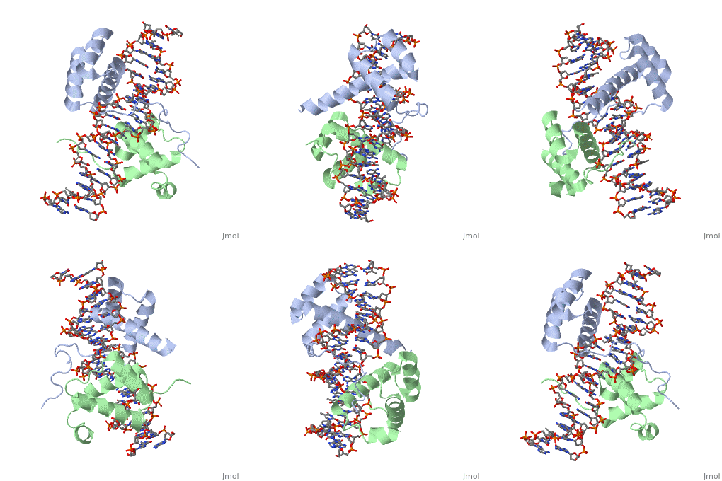
Tyrone Evans Hox Proteins sandbox
From Proteopedia
(Difference between revisions)
| (3 intermediate revisions not shown.) | |||
| Line 18: | Line 18: | ||
== Interaction with DNA == | == Interaction with DNA == | ||
| - | A homeodomain is an essential part that all Hox proteins possess. The homeodomain can be found within the homeobox, a DNA sequence found within genes that are responsible of morphogenesis of living organisms. This sequence codes for homeodomain protein products, 60 amino-acid segments, which have specific folding patterns (helix-turn-helix) that allows them to bind with DNA through a <scene name='71/714951/Taaat_sequence/1'>5’-TAAAT-3’</scene> core motif. This domain | + | A homeodomain is an essential part that all Hox proteins possess. The homeodomain can be found within the homeobox, a DNA sequence found within genes that are responsible of morphogenesis of living organisms. This sequence codes for homeodomain protein products, 60 amino-acid segments, which have specific folding patterns (helix-turn-helix) that allows them to bind with DNA through a <scene name='71/714951/Taaat_sequence/1'>5’-TAAAT-3’</scene> core motif. This domain consists of three helical regions folded into a tight spherical structure. There are <scene name='71/714951/Both_n_and_c-terminal_helices/1'>two antiparallel N-terminal helices and one C-terminal helix</scene> (Recognition helix) within this domain. The C-terminal helix binds directly with DNA through the use of <scene name='71/714951/H_bond_between_gln_and_p/1'>Hydrogen bonds</scene> and <scene name='71/714951/Hydrophobic_interaction/3'>Hydrophobic interactions</scene> between side chains and the outer bases and thymine methyl groups within the major groove of a DNA helix. Ionic bonding can also be observed between the bases of the DNA sequence and the amino acids of the protein dimers. A more specific <scene name='71/714951/Ionic_bond/1'>Ionic interaction</scene> is the one between LYS 207 and an oxygen atom of a phosphate group. The <scene name='71/714951/Recognition_helix/1'>Recognition helix</scene> within homeodomain binds within the <scene name='71/714951/Dna_major_groove/1'>major groove</scene> of a DNA helix, while the <scene name='71/714951/Amino-terminal_tail/1'>Amino-Terminal tail</scene> binds within the <scene name='71/714951/Dna_minor_groove/1'>minor groove</scene> of a DNA helix<ref>"Hox Gene." Wikipedia. Wikimedia Foundation, 14 Aug. 2015. Web. 20 Oct. 2015.</ref>. |
== Human Hox Genes<ref>"Hox Gene." Wikipedia. Wikimedia Foundation, 14 Aug. 2015. Web. 20 Oct. 2015.</ref> == | == Human Hox Genes<ref>"Hox Gene." Wikipedia. Wikimedia Foundation, 14 Aug. 2015. Web. 20 Oct. 2015.</ref> == | ||
| Line 32: | Line 32: | ||
== Structural highlights<ref>Berman, Helen M., John Westbrook, Zukang Feng, Gary Gilliland, T. N. Bhat, Helge Weissig, Ilya N. Shindyalov, and Philip E. Bourne. "The Protein Data Bank." RCSB. Nucl. Acids Res. (2000) 28 (1): 235-242 Web.</ref> == | == Structural highlights<ref>Berman, Helen M., John Westbrook, Zukang Feng, Gary Gilliland, T. N. Bhat, Helge Weissig, Ilya N. Shindyalov, and Philip E. Bourne. "The Protein Data Bank." RCSB. Nucl. Acids Res. (2000) 28 (1): 235-242 Web.</ref> == | ||
<scene name='71/714951/Taaat_sequence/1'>5’-TAAT-3’</scene> | <scene name='71/714951/Taaat_sequence/1'>5’-TAAT-3’</scene> | ||
| + | |||
| + | <scene name='71/714951/Both_n_and_c-terminal_helices/1'>Two antiparallel N-terminal helices and one C-terminal helix</scene> | ||
<scene name='71/714951/H_bond_between_gln_and_p/1'>Hydrogen bonds</scene> | <scene name='71/714951/H_bond_between_gln_and_p/1'>Hydrogen bonds</scene> | ||
Current revision
Hox Proteins
| |||||||||||

