101d
From Proteopedia
(Difference between revisions)
| (12 intermediate revisions not shown.) | |||
| Line 1: | Line 1: | ||
| - | [[Image:101d.gif|left|200px]] | ||
| - | + | ==REFINEMENT OF NETROPSIN BOUND TO DNA: BIAS AND FEEDBACK IN ELECTRON DENSITY MAP INTERPRETATION== | |
| - | + | <StructureSection load='101d' size='340' side='right'caption='[[101d]], [[Resolution|resolution]] 2.25Å' scene=''> | |
| - | + | == Structural highlights == | |
| - | + | <table><tr><td colspan='2'>[[101d]] is a 2 chain structure. Full crystallographic information is available from [http://oca.weizmann.ac.il/oca-bin/ocashort?id=101D OCA]. For a <b>guided tour on the structure components</b> use [https://proteopedia.org/fgij/fg.htm?mol=101D FirstGlance]. <br> | |
| - | + | </td></tr><tr id='method'><td class="sblockLbl"><b>[[Empirical_models|Method:]]</b></td><td class="sblockDat" id="methodDat">X-ray diffraction, [[Resolution|Resolution]] 2.25Å</td></tr> | |
| - | + | <tr id='ligand'><td class="sblockLbl"><b>[[Ligand|Ligands:]]</b></td><td class="sblockDat" id="ligandDat"><scene name='pdbligand=CBR:5-BROMO-2-DEOXY-CYTIDINE-5-MONOPHOSPHATE'>CBR</scene>, <scene name='pdbligand=MG:MAGNESIUM+ION'>MG</scene>, <scene name='pdbligand=NT:NETROPSIN'>NT</scene></td></tr> | |
| - | + | <tr id='resources'><td class="sblockLbl"><b>Resources:</b></td><td class="sblockDat"><span class='plainlinks'>[https://proteopedia.org/fgij/fg.htm?mol=101d FirstGlance], [http://oca.weizmann.ac.il/oca-bin/ocaids?id=101d OCA], [https://pdbe.org/101d PDBe], [https://www.rcsb.org/pdb/explore.do?structureId=101d RCSB], [https://www.ebi.ac.uk/pdbsum/101d PDBsum], [https://prosat.h-its.org/prosat/prosatexe?pdbcode=101d ProSAT]</span></td></tr> | |
| - | + | </table> | |
| - | + | <div style="background-color:#fffaf0;"> | |
| - | + | == Publication Abstract from PubMed == | |
| - | + | ||
| - | + | ||
| - | + | ||
| - | + | ||
| - | == | + | |
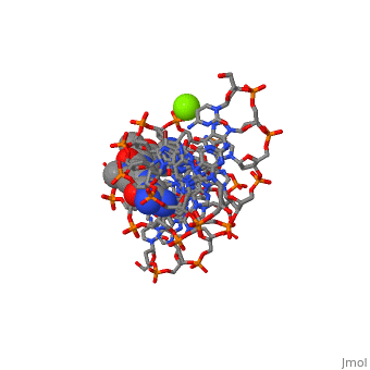
The X-ray crystal structure of the complex of the B-DNA dodecamer CGCGAATTCGCG with the antitumor drug netropsin has been reexamined to locate the drug accurately for computer-based drug design. The optimum solution is with the drug centered in the AATT region of the minor groove, making three good bifurcated hydrogen bonds with adenine N3 and thymine O2 atoms along the floor of the groove. Pyrrole rings of netropsin are packed against the C2 positions of adenines, leaving no room for the amine group of guanine and, hence, providing a structural rationale for the A.T specificity of netropsin. An alternative positioning in which the drug is shifted along the minor groove by ca. one-half base pair step is rejected on the basis of free R factor calculations and the appearance of the original drug-free difference maps. Final omit maps, although of more pleasing appearance, are not a dependable means of discriminating between right and wrong structures. The shifted alternative drug position ignores potential hydrogen bonding along the floor of the groove, provides no explanation for netropsin's observed A.T specificity, and is contradicted by NMR results [Patel, D. J. (1982) Proc. Natl. Acad. Sci. U.S.A. 79, 6424]. | The X-ray crystal structure of the complex of the B-DNA dodecamer CGCGAATTCGCG with the antitumor drug netropsin has been reexamined to locate the drug accurately for computer-based drug design. The optimum solution is with the drug centered in the AATT region of the minor groove, making three good bifurcated hydrogen bonds with adenine N3 and thymine O2 atoms along the floor of the groove. Pyrrole rings of netropsin are packed against the C2 positions of adenines, leaving no room for the amine group of guanine and, hence, providing a structural rationale for the A.T specificity of netropsin. An alternative positioning in which the drug is shifted along the minor groove by ca. one-half base pair step is rejected on the basis of free R factor calculations and the appearance of the original drug-free difference maps. Final omit maps, although of more pleasing appearance, are not a dependable means of discriminating between right and wrong structures. The shifted alternative drug position ignores potential hydrogen bonding along the floor of the groove, provides no explanation for netropsin's observed A.T specificity, and is contradicted by NMR results [Patel, D. J. (1982) Proc. Natl. Acad. Sci. U.S.A. 79, 6424]. | ||
| - | + | Refinement of netropsin bound to DNA: bias and feedback in electron density map interpretation.,Goodsell DS, Kopka ML, Dickerson RE Biochemistry. 1995 Apr 18;34(15):4983-93. PMID:7711020<ref>PMID:7711020</ref> | |
| - | + | ||
| - | + | ||
| - | + | ||
| - | Refinement of netropsin bound to DNA: bias and feedback in electron density map interpretation., Goodsell DS, Kopka ML, Dickerson RE | + | |
| - | + | ||
| - | + | ||
| - | + | ||
| - | + | ||
| - | + | ||
| - | + | ||
| - | + | ||
| - | + | ||
| - | + | From MEDLINE®/PubMed®, a database of the U.S. National Library of Medicine.<br> | |
| + | </div> | ||
| + | <div class="pdbe-citations 101d" style="background-color:#fffaf0;"></div> | ||
| + | == References == | ||
| + | <references/> | ||
| + | __TOC__ | ||
| + | </StructureSection> | ||
| + | [[Category: Large Structures]] | ||
| + | [[Category: Dickerson RE]] | ||
| + | [[Category: Goodsell DS]] | ||
| + | [[Category: Kopka ML]] | ||
Current revision
REFINEMENT OF NETROPSIN BOUND TO DNA: BIAS AND FEEDBACK IN ELECTRON DENSITY MAP INTERPRETATION
| |||||||||||