Actin
From Proteopedia
(Difference between revisions)
| (14 intermediate revisions not shown.) | |||
| Line 4: | Line 4: | ||
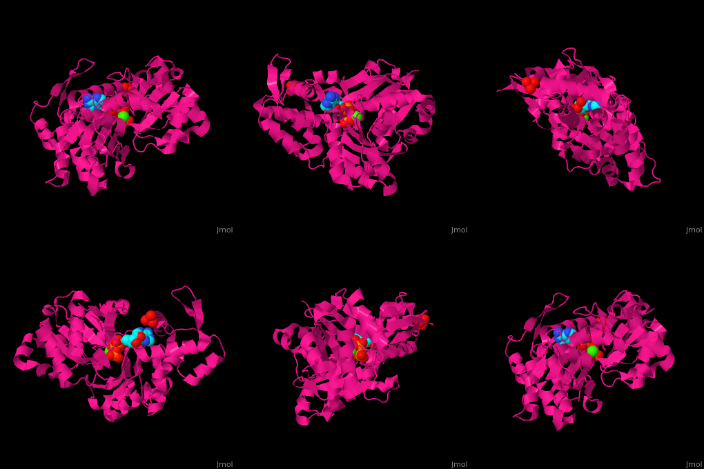
[[Actin]] is a protein found in all eukaryotic cells.<ref>PMID:11474115</ref> It is the monomer of two types of filaments: microfilaments which are part of the cytoskeleton and thin filaments which are part of muscles. Three isoforms of actin are identified: α (Aa) (or G-actin) found in muscle tissue, β (Ab) and γ (Ag) actins are components of the cytoskeleton. F-actin is Aa bound to ATP. For more details see:<br /> *[[F-actin]]<br /> *[[Non-polymerizable monomeric actin]].<br /> <scene name='Actin/Cv/2'>Click here to see the difference between 2 conformations of bovine Ag actin</scene> (PDB entries [[1hlu]] and [[2btf]]; morph was taken from [http://molmovdb.org/cgi-bin/movie.cgi Gallery of Morphs] of the [http://molmovdb.org Yale Morph Server]). Actin participates in muscle contraction, cell motility, cell division and cytokinesis. Actin associated with myosin is responsible for most cell movements. | [[Actin]] is a protein found in all eukaryotic cells.<ref>PMID:11474115</ref> It is the monomer of two types of filaments: microfilaments which are part of the cytoskeleton and thin filaments which are part of muscles. Three isoforms of actin are identified: α (Aa) (or G-actin) found in muscle tissue, β (Ab) and γ (Ag) actins are components of the cytoskeleton. F-actin is Aa bound to ATP. For more details see:<br /> *[[F-actin]]<br /> *[[Non-polymerizable monomeric actin]].<br /> <scene name='Actin/Cv/2'>Click here to see the difference between 2 conformations of bovine Ag actin</scene> (PDB entries [[1hlu]] and [[2btf]]; morph was taken from [http://molmovdb.org/cgi-bin/movie.cgi Gallery of Morphs] of the [http://molmovdb.org Yale Morph Server]). Actin participates in muscle contraction, cell motility, cell division and cytokinesis. Actin associated with myosin is responsible for most cell movements. | ||
| + | *'''α actin''' is found exclusively in muscle fibres.<br /> | ||
| + | *'''β actin''' is required for early embryonic development<ref>PMID:21900491</ref>.<br /> | ||
| + | *'''γ actin''' is required for cytoskeletal maintenance<ref>PMID:19497859</ref>. | ||
| + | |||
| + | See also [[Actin Protein (Hebrew)]] | ||
== Disease == | == Disease == | ||
| Line 11: | Line 16: | ||
== Structural highlights == | == Structural highlights == | ||
| - | <scene name='43/430015/Cv/ | + | <scene name='43/430015/Cv/10'>Actin binds ATP</scene> in a cleft. Water molecules are shown as red spheres. <scene name='43/430015/Cv/13'>ATP and Ca2+ ion are located in cleft</scene>. <scene name='43/430015/Cv/11'>Click here to see Ca2+ ion coordination site</scene>.<ref>PMID:20540085</ref> It changes its conformation upon hydrolysis of its bound ATP to ADP. Actin filaments are polar. They are formed with all monomers having their clefts pointing in the same direction. |
| - | + | ||
== 3D Structures of Actin == | == 3D Structures of Actin == | ||
| + | [[Actin 3D structures]] | ||
| - | + | </StructureSection> | |
| - | + | ||
| - | + | ||
| - | + | ||
| - | + | ||
| - | + | ||
| - | + | ||
| - | + | ||
| - | + | ||
| - | + | ||
| - | + | ||
| - | + | ||
| - | + | ||
| - | + | ||
| - | + | ||
| - | + | ||
| - | + | ||
| - | + | ||
| - | + | ||
| - | + | ||
| - | + | ||
| - | + | ||
| - | + | ||
| - | + | ||
| - | + | ||
| - | + | ||
| - | + | ||
| - | + | ||
| - | + | ||
| - | + | ||
| - | + | ||
| - | + | ||
| - | + | ||
| - | + | ||
| - | + | ||
| - | + | ||
| - | + | ||
| - | + | ||
| - | + | ||
| - | + | ||
| - | + | ||
| - | + | ||
| - | + | ||
| - | + | ||
| - | + | ||
| - | + | ||
| - | + | ||
| - | + | ||
| - | + | ||
| - | + | ||
| - | + | ||
| - | + | ||
| - | + | ||
| - | + | ||
| - | + | ||
| - | + | ||
| - | + | ||
| - | + | ||
| - | + | ||
| - | + | ||
| - | + | ||
| - | + | ||
| - | + | ||
| - | + | ||
| - | + | ||
| - | + | ||
| - | + | ||
| - | + | ||
| - | + | ||
| - | + | ||
| - | + | ||
| - | + | ||
| - | + | ||
| - | + | ||
| - | + | ||
| - | + | ||
| - | + | ||
| - | **[[1atn]] – rAg+cDNase I<br /> | ||
| - | **[[3chw]] – Major DdAg+hProfilin+poly-Pro repeat – ''Dictyostelium discoideum''<br /> | ||
| - | **[[3ci5]], [[3cip]], [[1nmd]] - Major DdAg+hGelsolin<br /> | ||
| - | **[[3a5l]], [[3a5m]], [[3a5n]], [[3a5o]] - Major DdAg+hGelsolin (mutant)<br /> | ||
| - | **[[1c0g]], [[1dej]] – DdAg:tetrahymenatA+hGelsolin<br /> | ||
| - | **[[1nm1]] - DdAg+hGelsolin+Mg-ATP<br /> | ||
| - | **[[1nlv]], [[1c0f]] – DdAg+hGelsolin<br /> | ||
| - | **[[1d4x]] – Ag 1/3+hGelsolin – ''C. elegans''<br /> | ||
| - | **[[1h1v]] – hAg+rGelsolin<br /> | ||
| - | **[[1hlu]], [[2btf]] – cAg+cProfilin<br /> | ||
| - | **[[1o1g]], [[1o1a]], [[1o1b]], [[1o1c]], [[1o1d]], [[1o1e]], [[1o1f]], [[1o18]], [[1o19]], [[1mvw]], [[1m8q]] –cAg+cMyosin II - tomography<br /> | ||
| - | **[[3eks]] – DmAg 5c (mutant)<br /> | ||
| - | **[[3eku]] – DmAg 5c (mutant)+cytochalasin D<br /> | ||
| - | **[[3el2]], [[2hf4]] - DmAg 5c (mutant)+Ca-ATP<br /> | ||
| - | **[[2hf3]] - DmAg 5c (mutant)+Ca-ADP<br /> | ||
| - | **[[3w3d]] – cAg + DNase I<br /> | ||
| - | **[[3b63]] – actin filament – ''Limulus polyphemus''<br /> | ||
| - | **[[1yvn]] – yA (mutant)+hGelsolin <br /> | ||
| - | **[[1yag]] - yA+hGelsolin<br /> | ||
| - | **[[3mmv]] – DmA-5C+spire WH2 domain<br /> | ||
| - | **[[3mn6]], [[3mn7]], [[3mn9]] - DmA-5C (mutant)+spire<br /> | ||
| - | **[[4efh]] – A + spire domains C,D – ''Acanthamoeba castellanii'' | ||
| - | }} | ||
== Reference == | == Reference == | ||
<references/> | <references/> | ||
[[Category:Topic Page]] | [[Category:Topic Page]] | ||
Current revision
| |||||||||||
Reference
- ↑ Otterbein LR, Graceffa P, Dominguez R. The crystal structure of uncomplexed actin in the ADP state. Science. 2001 Jul 27;293(5530):708-11. PMID:11474115 doi:10.1126/science.1059700
- ↑ Bunnell TM, Burbach BJ, Shimizu Y, Ervasti JM. β-Actin specifically controls cell growth, migration, and the G-actin pool. Mol Biol Cell. 2011 Nov;22(21):4047-58. PMID:21900491 doi:10.1091/mbc.E11-06-0582
- ↑ Belyantseva IA, Perrin BJ, Sonnemann KJ, Zhu M, Stepanyan R, McGee J, Frolenkov GI, Walsh EJ, Friderici KH, Friedman TB, Ervasti JM. Gamma-actin is required for cytoskeletal maintenance but not development. Proc Natl Acad Sci U S A. 2009 Jun 16;106(24):9703-8. PMID:19497859 doi:10.1073/pnas.0900221106
- ↑ Wang H, Robinson RC, Burtnick LD. The structure of native G-actin. Cytoskeleton (Hoboken). 2010 Jul;67(7):456-65. PMID:20540085 doi:10.1002/cm.20458

