This old version of Proteopedia is provided for student assignments while the new version is undergoing repairs. Content and edits done in this old version of Proteopedia after March 1, 2026 will eventually be lost when it is retired in about June of 2026.
Apply for new accounts at the new Proteopedia. Your logins will work in both the old and new versions.
1cbs
From Proteopedia
(Difference between revisions)
| (13 intermediate revisions not shown.) | |||
| Line 1: | Line 1: | ||
| - | [[Image:1cbs.gif|left|200px]] | ||
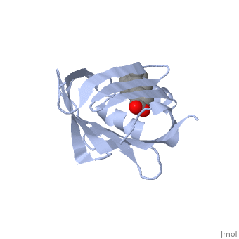
| - | + | ==CRYSTAL STRUCTURE OF CELLULAR RETINOIC-ACID-BINDING PROTEINS I AND II IN COMPLEX WITH ALL-TRANS-RETINOIC ACID AND A SYNTHETIC RETINOID== | |
| - | + | <StructureSection load='1cbs' size='340' side='right'caption='[[1cbs]], [[Resolution|resolution]] 1.80Å' scene=''> | |
| - | + | == Structural highlights == | |
| - | | | + | <table><tr><td colspan='2'>[[1cbs]] is a 1 chain structure with sequence from [https://en.wikipedia.org/wiki/Homo_sapiens Homo sapiens]. Full crystallographic information is available from [http://oca.weizmann.ac.il/oca-bin/ocashort?id=1CBS OCA]. For a <b>guided tour on the structure components</b> use [https://proteopedia.org/fgij/fg.htm?mol=1CBS FirstGlance]. <br> |
| - | + | </td></tr><tr id='method'><td class="sblockLbl"><b>[[Empirical_models|Method:]]</b></td><td class="sblockDat" id="methodDat">X-ray diffraction, [[Resolution|Resolution]] 1.8Å</td></tr> | |
| - | + | <tr id='ligand'><td class="sblockLbl"><b>[[Ligand|Ligands:]]</b></td><td class="sblockDat" id="ligandDat"><scene name='pdbligand=REA:RETINOIC+ACID'>REA</scene></td></tr> | |
| - | + | <tr id='resources'><td class="sblockLbl"><b>Resources:</b></td><td class="sblockDat"><span class='plainlinks'>[https://proteopedia.org/fgij/fg.htm?mol=1cbs FirstGlance], [http://oca.weizmann.ac.il/oca-bin/ocaids?id=1cbs OCA], [https://pdbe.org/1cbs PDBe], [https://www.rcsb.org/pdb/explore.do?structureId=1cbs RCSB], [https://www.ebi.ac.uk/pdbsum/1cbs PDBsum], [https://prosat.h-its.org/prosat/prosatexe?pdbcode=1cbs ProSAT]</span></td></tr> | |
| - | + | </table> | |
| - | + | == Function == | |
| - | + | [https://www.uniprot.org/uniprot/RABP2_HUMAN RABP2_HUMAN] Transports retinoic acid to the nucleus. Regulates the access of retinoic acid to the nuclear retinoic acid receptors. | |
| + | == Evolutionary Conservation == | ||
| + | [[Image:Consurf_key_small.gif|200px|right]] | ||
| + | Check<jmol> | ||
| + | <jmolCheckbox> | ||
| + | <scriptWhenChecked>; select protein; define ~consurf_to_do selected; consurf_initial_scene = true; script "/wiki/ConSurf/cb/1cbs_consurf.spt"</scriptWhenChecked> | ||
| + | <scriptWhenUnchecked>script /wiki/extensions/Proteopedia/spt/initialview01.spt</scriptWhenUnchecked> | ||
| + | <text>to colour the structure by Evolutionary Conservation</text> | ||
| + | </jmolCheckbox> | ||
| + | </jmol>, as determined by [http://consurfdb.tau.ac.il/ ConSurfDB]. You may read the [[Conservation%2C_Evolutionary|explanation]] of the method and the full data available from [http://bental.tau.ac.il/new_ConSurfDB/main_output.php?pdb_ID=1cbs ConSurf]. | ||
| + | <div style="clear:both"></div> | ||
| - | + | ==See Also== | |
| - | + | *[[CRABP I ( Cellular Retinoic Acid Binding Protein )|CRABP I ( Cellular Retinoic Acid Binding Protein )]] | |
| - | + | *[[Cellular retinoic acid-binding protein|Cellular retinoic acid-binding protein]] | |
| - | == | + | *[[Cellular retinoic acid-binding protein 3D structures|Cellular retinoic acid-binding protein 3D structures]] |
| - | + | *[[Gustavo Elberto Epalza Sanchez/Sandbox 1|Gustavo Elberto Epalza Sanchez/Sandbox 1]] | |
| - | + | *[[Molecular Playground/CRABP I (Cellular Retinoic Acid Binding Protein)|Molecular Playground/CRABP I (Cellular Retinoic Acid Binding Protein)]] | |
| - | + | __TOC__ | |
| - | + | </StructureSection> | |
| - | + | ||
| - | + | ||
| - | + | ||
[[Category: Homo sapiens]] | [[Category: Homo sapiens]] | ||
| - | [[Category: | + | [[Category: Large Structures]] |
| - | [[Category: Bergfors | + | [[Category: Bergfors T]] |
| - | [[Category: Jones | + | [[Category: Jones TA]] |
| - | [[Category: Kleywegt | + | [[Category: Kleywegt GJ]] |
| - | + | ||
| - | + | ||
| - | + | ||
Current revision
CRYSTAL STRUCTURE OF CELLULAR RETINOIC-ACID-BINDING PROTEINS I AND II IN COMPLEX WITH ALL-TRANS-RETINOIC ACID AND A SYNTHETIC RETINOID
| |||||||||||