|
|
| (15 intermediate revisions not shown.) |
| Line 1: |
Line 1: |
| - | [[Image:1dsn.gif|left|200px]] | |
| | | | |
| - | {{Structure
| + | ==D60S N-TERMINAL LOBE HUMAN LACTOFERRIN== |
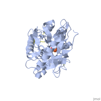
| - | |PDB= 1dsn |SIZE=350|CAPTION= <scene name='initialview01'>1dsn</scene>, resolution 2.05Å
| + | <StructureSection load='1dsn' size='340' side='right'caption='[[1dsn]], [[Resolution|resolution]] 2.05Å' scene=''> |
| - | |SITE=
| + | == Structural highlights == |
| - | |LIGAND= <scene name='pdbligand=CO3:CARBONATE+ION'>CO3</scene>, <scene name='pdbligand=FE:FE+(III)+ION'>FE</scene> | + | <table><tr><td colspan='2'>[[1dsn]] is a 1 chain structure with sequence from [https://en.wikipedia.org/wiki/Homo_sapiens Homo sapiens]. Full crystallographic information is available from [http://oca.weizmann.ac.il/oca-bin/ocashort?id=1DSN OCA]. For a <b>guided tour on the structure components</b> use [https://proteopedia.org/fgij/fg.htm?mol=1DSN FirstGlance]. <br> |
| - | |ACTIVITY=
| + | </td></tr><tr id='method'><td class="sblockLbl"><b>[[Empirical_models|Method:]]</b></td><td class="sblockDat" id="methodDat">X-ray diffraction, [[Resolution|Resolution]] 2.05Å</td></tr> |
| - | |GENE= LFN ([http://www.ncbi.nlm.nih.gov/Taxonomy/Browser/wwwtax.cgi?mode=Info&srchmode=5&id=9606 Homo sapiens])
| + | <tr id='ligand'><td class="sblockLbl"><b>[[Ligand|Ligands:]]</b></td><td class="sblockDat" id="ligandDat"><scene name='pdbligand=CO3:CARBONATE+ION'>CO3</scene>, <scene name='pdbligand=FE:FE+(III)+ION'>FE</scene></td></tr> |
| - | |DOMAIN=
| + | <tr id='resources'><td class="sblockLbl"><b>Resources:</b></td><td class="sblockDat"><span class='plainlinks'>[https://proteopedia.org/fgij/fg.htm?mol=1dsn FirstGlance], [http://oca.weizmann.ac.il/oca-bin/ocaids?id=1dsn OCA], [https://pdbe.org/1dsn PDBe], [https://www.rcsb.org/pdb/explore.do?structureId=1dsn RCSB], [https://www.ebi.ac.uk/pdbsum/1dsn PDBsum], [https://prosat.h-its.org/prosat/prosatexe?pdbcode=1dsn ProSAT]</span></td></tr> |
| - | |RELATEDENTRY=
| + | </table> |
| - | |RESOURCES=<span class='plainlinks'>[http://oca.weizmann.ac.il/oca-docs/fgij/fg.htm?mol=1dsn FirstGlance], [http://oca.weizmann.ac.il/oca-bin/ocaids?id=1dsn OCA], [http://www.ebi.ac.uk/pdbsum/1dsn PDBsum], [http://www.rcsb.org/pdb/explore.do?structureId=1dsn RCSB]</span>
| + | == Function == |
| - | }}
| + | [https://www.uniprot.org/uniprot/TRFL_HUMAN TRFL_HUMAN] Transferrins are iron binding transport proteins which can bind two Fe(3+) ions in association with the binding of an anion, usually bicarbonate.<ref>PMID:12535064</ref> <ref>PMID:22320386</ref> Lactotransferrin has antimicrobial activity which depends on the extracellular cation concentration.<ref>PMID:12535064</ref> <ref>PMID:22320386</ref> Lactoferroxins A, B and C have opioid antagonist activity. Lactoferroxin A shows preference for mu-receptors, while lactoferroxin B and C have somewhat higher degrees of preference for kappa-receptors than for mu-receptors.<ref>PMID:12535064</ref> <ref>PMID:22320386</ref> The lactotransferrin transferrin-like domain 1 functions as a serine protease of the peptidase S60 family that cuts arginine rich regions. This function contributes to the antimicrobial activity.<ref>PMID:12535064</ref> <ref>PMID:22320386</ref> Isoform DeltaLf: transcription factor with antiproliferative properties and inducing cell cycle arrest. Binds to DeltaLf response element found in the SKP1, BAX, DCPS, and SELH promoters.<ref>PMID:12535064</ref> <ref>PMID:22320386</ref> |
| | + | == Evolutionary Conservation == |
| | + | [[Image:Consurf_key_small.gif|200px|right]] |
| | + | Check<jmol> |
| | + | <jmolCheckbox> |
| | + | <scriptWhenChecked>; select protein; define ~consurf_to_do selected; consurf_initial_scene = true; script "/wiki/ConSurf/ds/1dsn_consurf.spt"</scriptWhenChecked> |
| | + | <scriptWhenUnchecked>script /wiki/extensions/Proteopedia/spt/initialview01.spt</scriptWhenUnchecked> |
| | + | <text>to colour the structure by Evolutionary Conservation</text> |
| | + | </jmolCheckbox> |
| | + | </jmol>, as determined by [http://consurfdb.tau.ac.il/ ConSurfDB]. You may read the [[Conservation%2C_Evolutionary|explanation]] of the method and the full data available from [http://bental.tau.ac.il/new_ConSurfDB/main_output.php?pdb_ID=1dsn ConSurf]. |
| | + | <div style="clear:both"></div> |
| | | | |
| - | '''D60S N-TERMINAL LOBE HUMAN LACTOFERRIN'''
| + | ==See Also== |
| - | | + | *[[Lactoferrin|Lactoferrin]] |
| - | | + | == References == |
| - | ==Overview== | + | <references/> |
| - | The crystal structure of a site-specific mutant of the N-terminal half-molecule of human lactoferrin, Lf(N), in which the iron ligand Asp60 has been mutated to Ser, has been determined at 2.05 A resolution in order to determine the effects of the mutation on iron binding and domain closure. Yellow monoclinic crystals of the D60S mutant, in its iron-bound form, were prepared, and have unit cell dimensions a = 110.2 A, b = 57.0 A, c = 55.2 A, beta = 97.6 degrees, space group C2, with one molecule of 333 residues in the asymmetric unit. The structure was determined by molecular replacement, using the wild-type Lf(N) as search model, and was refined by restrained least-squares methods. The final model, comprising 2451 protein atoms (from residues 2 to 315) one Fe3+ and one CO2-(3), and 107 water molecules, gives an R-factor of 0.175 for all data in the resolution range 20.0 to 2.05 A. The model conforms well with standard geometry, having root-mean-square deviations of 0.014 A and 1.2 degrees from standard bond lengths and angles. The structure of the D60S mutant deviates in two important respects from the parent Lf(N) molecule. At the mutation site the Ser side-chain neither binds to the iron atom nor makes any interdomain contact as the substituted Asp does; instead a water molecule fills the iron coordination site and participates in interdomain hydrogen bonding. The domain closure is also changed, with the D60S mutant having a more closed conformation. Consideration of crystal packing suggests that the altered domain closure is a genuine molecular property but both the iron coordination and interdomain contacts are consistent with weakened iron binding in the mutant. The implications for iron binding in transferrins generally are discussed.
| + | __TOC__ |
| - | | + | </StructureSection> |
| - | ==About this Structure==
| + | |
| - | 1DSN is a [[Single protein]] structure of sequence from [http://en.wikipedia.org/wiki/Homo_sapiens Homo sapiens]. Full crystallographic information is available from [http://oca.weizmann.ac.il/oca-bin/ocashort?id=1DSN OCA].
| + | |
| - | | + | |
| - | ==Reference== | + | |
| - | Altered domain closure and iron binding in transferrins: the crystal structure of the Asp60Ser mutant of the amino-terminal half-molecule of human lactoferrin., Faber HR, Bland T, Day CL, Norris GE, Tweedie JW, Baker EN, J Mol Biol. 1996 Feb 23;256(2):352-63. PMID:[http://www.ncbi.nlm.nih.gov/pubmed/8594202 8594202]
| + | |
| | [[Category: Homo sapiens]] | | [[Category: Homo sapiens]] |
| - | [[Category: Single protein]] | + | [[Category: Large Structures]] |
| - | [[Category: Baker, E N.]] | + | [[Category: Baker EN]] |
| - | [[Category: Faber, H R.]] | + | [[Category: Faber HR]] |
| - | [[Category: Norris, G E.]] | + | [[Category: Norris GE]] |
| - | [[Category: glycoprotein]]
| + | |
| - | [[Category: iron transport]]
| + | |
| - | [[Category: metal-binding]]
| + | |
| - | | + | |
| - | ''Page seeded by [http://oca.weizmann.ac.il/oca OCA ] on Sun Mar 30 19:47:53 2008''
| + | |
| Structural highlights
Function
TRFL_HUMAN Transferrins are iron binding transport proteins which can bind two Fe(3+) ions in association with the binding of an anion, usually bicarbonate.[1] [2] Lactotransferrin has antimicrobial activity which depends on the extracellular cation concentration.[3] [4] Lactoferroxins A, B and C have opioid antagonist activity. Lactoferroxin A shows preference for mu-receptors, while lactoferroxin B and C have somewhat higher degrees of preference for kappa-receptors than for mu-receptors.[5] [6] The lactotransferrin transferrin-like domain 1 functions as a serine protease of the peptidase S60 family that cuts arginine rich regions. This function contributes to the antimicrobial activity.[7] [8] Isoform DeltaLf: transcription factor with antiproliferative properties and inducing cell cycle arrest. Binds to DeltaLf response element found in the SKP1, BAX, DCPS, and SELH promoters.[9] [10]
Evolutionary Conservation
Check, as determined by ConSurfDB. You may read the explanation of the method and the full data available from ConSurf.
See Also
References
- ↑ Hendrixson DR, Qiu J, Shewry SC, Fink DL, Petty S, Baker EN, Plaut AG, St Geme JW 3rd. Human milk lactoferrin is a serine protease that cleaves Haemophilus surface proteins at arginine-rich sites. Mol Microbiol. 2003 Feb;47(3):607-17. PMID:12535064
- ↑ Mariller C, Hardiville S, Hoedt E, Huvent I, Pina-Canseco S, Pierce A. Delta-lactoferrin, an intracellular lactoferrin isoform that acts as a transcription factor. Biochem Cell Biol. 2012 Jun;90(3):307-19. doi: 10.1139/o11-070. Epub 2012 Feb 9. PMID:22320386 doi:http://dx.doi.org/10.1139/o11-070
|