Sandbox Reserved 431
From Proteopedia
| (11 intermediate revisions not shown.) | |||
| Line 7: | Line 7: | ||
[[Student Projects for UMass Chemistry 423 Spring 2016]] | [[Student Projects for UMass Chemistry 423 Spring 2016]] | ||
| - | <StructureSection load='3c6g' size='350' side='right' caption='caption for Molecular Playground (PDB entry [[3c6g]])' scene=''> | ||
==Introduction== | ==Introduction== | ||
| - | Rickets is a disease | + | Rickets is a disease resulting from prolonged vitamin D deficiency. As vitamin D is vital for the absorption of phosphorus and calcium, a deficiency would cause weakening of bones. Rickets also causes muscle weakness, skeletal deformities, dental problems, inhibition of growth, and muscle spasms. In some cases, rickets can be inherited due to mutations in genes responsible for coding for human CYP2R1. |
| - | + | ||
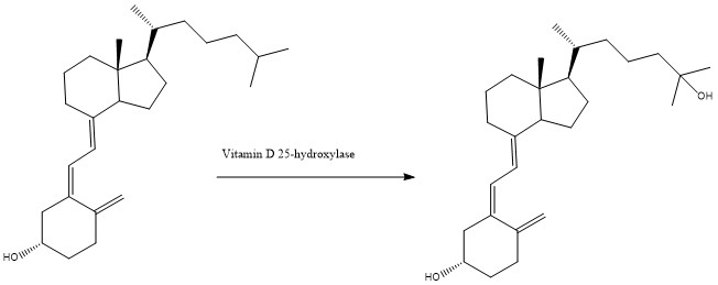
| + | In the human body, CYP2R1, a member of the cytochrome P450 family, is responsible for the first steps of the conversion of vitamin D into a bioavailable form within the liver. CYP2R1 is also known as Vitamin D 25-hydroxylase as it hydroxylates vitamin D3 into calcidiol, the bioavailable form of the vitamin, which would then be converted to calcitriol via the enzyme 25-hydroxyvitamin D3 1-alpha-hydroxylase, as seen in figure 1. | ||
| + | |||
| + | [[Image:Action_Of_CYP2R1.jpg]] | ||
| + | |||
| + | Fig. 1, the conversion of vitamin D3 into calcidiol via Vitamin D 25-hydroxylase | ||
| + | |||
| + | <scene name='48/483888/Human_p450/1'>Human P450 Cytochrome</scene> is shown with amino acids in teal and the heme center shown as a space filling model, with the nitrogen shown in blue, carbon in gray, oxygen in red, and the iron center shown in orange | ||
| + | |||
| + | <StructureSection load='3c6g' size='350' side='right' caption='Structure of Human Cytochrome p450' scene=''> | ||
==Overall Structure== | ==Overall Structure== | ||
| - | Cytochrome P450 | + | Cytochrome P450 is an <scene name='48/483888/Cytochrome_p450_dimer/1'>asymmetric dimer</scene>, which means the protein is made of two subunits that are structurally very similar to one another, but not identical. Each dimeric subunit contain 12 α-helices (labeled A-L) along with some β-sheets that are localized to one side of the molecule. Helices F and G from each of the units form the dimer interface of cytochrome P450, and are involved in the formation of the active site. This dimeric interface of the protein is stabilized by <scene name='48/483888/Cytochrome_p450_interface/1'>hydrogen bonding interactions</scene> between residues from the G helix of one the units with residues located on the the F helix of the second unit, and vice versa. Two molecules of 2-hydroxypropyl-β-cyclodextrin are also found near the dimer interface. The cyclodextrins are believed to help further stabilize the protein, and also shield the hydrophobic part of the F-G helix transition loop from the solvent by <scene name='48/483888/Cytochrome_p450_cyclodextrins/1'>encapsulating the Phe240 residue within its cavity</scene>. Cytochrome P450 has an apparent mass of ~120 kDa. |
==Binding Interactions== | ==Binding Interactions== | ||
| Line 30: | Line 37: | ||
<scene name='48/483888/Hemegroup/1'>Leu99 and Arg445</scene> | <scene name='48/483888/Hemegroup/1'>Leu99 and Arg445</scene> | ||
| - | + | </StructureSection> | |
| - | + | ||
==Quiz Question 1== | ==Quiz Question 1== | ||
| - | + | 1A) Why is Proline so poorly suited for inclusion in Alpha Helices? | |
| - | + | A) It cannot hydrogen bond due to the position of its amide | |
| + | B) The residue is incapable of forming the correct phi and psi angles in a helix | ||
| + | C) The steric hindrance of its sidechain | ||
| + | D) A and C | ||
| + | E) All of the above | ||
| - | + | 1B) This protein being a dimer, has symmetry between its two large sections, from <scene name='48/483888/Gandfhelices/1'>this orientation</scene> where most of the molecule has been cut away for simplicity, you can see where one half (in green) comes within close proximity of the other half (in blue). These 2 pairs of helices help hold the dimer together via electrostatic interactions. If the black residue is Arg, and the white residue is Asp, what is most likely to be on the opposite helix | |
| + | (Arg match, Asp match) | ||
| + | A) Asp, His | ||
| + | B) Gly, Val | ||
| + | C) Met, Lys | ||
==See Also== | ==See Also== | ||
Current revision
| This Sandbox is Reserved from January 19, 2016, through August 31, 2016 for use for Proteopedia Team Projects by the class Chemistry 423 Biochemistry for Chemists taught by Lynmarie K Thompson at University of Massachusetts Amherst, USA. This reservation includes Sandbox Reserved 425 through Sandbox Reserved 439. |
Contents |
Vitamin D activation by cytochrome P450, Rickets (3c6g)[1]
by Isabel Hand, Elizabeth Humble, Kati Johnson, Samantha Kriksceonaitis, and Matthew Tiller
Student Projects for UMass Chemistry 423 Spring 2016
Introduction
Rickets is a disease resulting from prolonged vitamin D deficiency. As vitamin D is vital for the absorption of phosphorus and calcium, a deficiency would cause weakening of bones. Rickets also causes muscle weakness, skeletal deformities, dental problems, inhibition of growth, and muscle spasms. In some cases, rickets can be inherited due to mutations in genes responsible for coding for human CYP2R1.
In the human body, CYP2R1, a member of the cytochrome P450 family, is responsible for the first steps of the conversion of vitamin D into a bioavailable form within the liver. CYP2R1 is also known as Vitamin D 25-hydroxylase as it hydroxylates vitamin D3 into calcidiol, the bioavailable form of the vitamin, which would then be converted to calcitriol via the enzyme 25-hydroxyvitamin D3 1-alpha-hydroxylase, as seen in figure 1.
Fig. 1, the conversion of vitamin D3 into calcidiol via Vitamin D 25-hydroxylase
is shown with amino acids in teal and the heme center shown as a space filling model, with the nitrogen shown in blue, carbon in gray, oxygen in red, and the iron center shown in orange
| |||||||||||
Quiz Question 1
1A) Why is Proline so poorly suited for inclusion in Alpha Helices?
    A) It cannot hydrogen bond due to the position of its amide
    B) The residue is incapable of forming the correct phi and psi angles in a helix
    C) The steric hindrance of its sidechain
    D) A and C
    E) All of the above
1B) This protein being a dimer, has symmetry between its two large sections, from where most of the molecule has been cut away for simplicity, you can see where one half (in green) comes within close proximity of the other half (in blue). These 2 pairs of helices help hold the dimer together via electrostatic interactions. If the black residue is Arg, and the white residue is Asp, what is most likely to be on the opposite helix
       (Arg match, Asp match)
    A) Asp, His
    B) Gly, Val
    C) Met, Lys
See Also
Credits
Introduction - Sami Kriksceonaitis
Overall Structure - Kati Johnson
Binding Interactions - Isabel Hand
Additional Features - Elizabeth Humble
Quiz Question 1 - Matthew Tiller
References
- ↑ Strushkevich N, Usanov SA, Plotnikov AN, Jones G, Park HW. Structural analysis of CYP2R1 in complex with vitamin D3. J Mol Biol. 2008 Jun 27;380(1):95-106. Epub 2008 Apr 8. PMID:18511070 doi:10.1016/j.jmb.2008.03.065

