Sandbox HEC
From Proteopedia
(Difference between revisions)
| (3 intermediate revisions not shown.) | |||
| Line 18: | Line 18: | ||
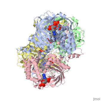
Human erythrocyte catalase is a negatively charged heme-containing monofunctional tetrameric enzyme that is prevalent among aerobic organisms <ref name= "Kodydková" >PMID:25152049</ref><ref name=Alfonso-Prietro>PMID:22516655</ref><ref name=Dash>PMID:22521743</ref><ref name=Diaz>PMID:22209752 </ref><ref name=Nishikawa>PMID:19385054 </ref>. The catalase fold contains an eight-sheeted anti-parallel beta-barrel domain linked to a six alpha-helical domain via a lengthy protein sequence. Residues within β1-β4 contribute to the heme variant, while residues within β5-β8 establish the NADPH binding site <ref name="Diaz" />. The positioning of the heme is determined by the proximal aromatic pyrrole compounds; in human erythrocyte catalase, catalytic His75 is <scene name='72/728053/Position_of_his75/3'>positioned</scene> above pyrrole ring III, further producing a His-III orientation and heme-b variant. The <scene name='72/728053/Active_site_1/2'>NADPH Binding site </scene> is located at the β,α-domain junction <ref name="Alfonso-Prietro" /><ref name="Diaz" />. When the NADPH molecule is bound, a right-handed clockwise helical formation is produced. In human erythrocyte catalase, only two of the four subunits allow for NADPH binding <ref name="Kodydková" /><ref name="Diaz" />. The active site contains a negatively charged tyrosine and a positively charged histidine situated, respectively, proximal and distal to the heme group. The histidine is responsible for the formation of Compound I during the first step of the catalase mechanism <ref name="Alfonso-Prietro" />. | Human erythrocyte catalase is a negatively charged heme-containing monofunctional tetrameric enzyme that is prevalent among aerobic organisms <ref name= "Kodydková" >PMID:25152049</ref><ref name=Alfonso-Prietro>PMID:22516655</ref><ref name=Dash>PMID:22521743</ref><ref name=Diaz>PMID:22209752 </ref><ref name=Nishikawa>PMID:19385054 </ref>. The catalase fold contains an eight-sheeted anti-parallel beta-barrel domain linked to a six alpha-helical domain via a lengthy protein sequence. Residues within β1-β4 contribute to the heme variant, while residues within β5-β8 establish the NADPH binding site <ref name="Diaz" />. The positioning of the heme is determined by the proximal aromatic pyrrole compounds; in human erythrocyte catalase, catalytic His75 is <scene name='72/728053/Position_of_his75/3'>positioned</scene> above pyrrole ring III, further producing a His-III orientation and heme-b variant. The <scene name='72/728053/Active_site_1/2'>NADPH Binding site </scene> is located at the β,α-domain junction <ref name="Alfonso-Prietro" /><ref name="Diaz" />. When the NADPH molecule is bound, a right-handed clockwise helical formation is produced. In human erythrocyte catalase, only two of the four subunits allow for NADPH binding <ref name="Kodydková" /><ref name="Diaz" />. The active site contains a negatively charged tyrosine and a positively charged histidine situated, respectively, proximal and distal to the heme group. The histidine is responsible for the formation of Compound I during the first step of the catalase mechanism <ref name="Alfonso-Prietro" />. | ||
| + | |||
| Line 23: | Line 24: | ||
[[Image:HEC mechanism.jpg.png|thumb|400px|'''Human Erythrocyte Catalase Mechanism''' This figure illustrates the two step mechanism of catalase. ]] | [[Image:HEC mechanism.jpg.png|thumb|400px|'''Human Erythrocyte Catalase Mechanism''' This figure illustrates the two step mechanism of catalase. ]] | ||
| - | == Mechanism == | ||
| + | |||
| + | |||
| + | |||
| + | |||
| + | |||
| + | == Mechanism == | ||
Stable forms of hydrogen peroxide are beneficial in biological reactions including hypoxia signal transduction, cell proliferation and differentiation regulation, as well as immune response mediation; however, it is toxic at high levels as free hydroxyl ions cannot be catalyzed by the body <ref name= Lennicke >PMID:26369938</ref>. Hydrogen peroxide acts to both the oxidizing and reducing agent of the iron. Catalase ultimately functions to break down hydrogen peroxide<ref name="Dash" />. This is accomplished in a two-step mechanism where the heme is first oxidized by a molecule of hydrogen peroxide to produce Compound I, a high energy oxyferryl cation radical intermediate, as well as a water molecule. Compound I is then immediately reduced by a second hydrogen peroxide molecule to produce a second molecule of water <ref name="Alfonso-Prietro" /><ref name="Diaz" />. The overall reaction results in two single-electron transfers from the iron atom of the heme group and the porphyrin from the oxoferryl radical, as well as a proton transfer from histidine. The mechanism is enthalpically driven by the distal histidine proton transfer as it is more exothermic than the electron transfers <ref name="Alfonso-Prietro" /><ref name="Diaz" /> . | Stable forms of hydrogen peroxide are beneficial in biological reactions including hypoxia signal transduction, cell proliferation and differentiation regulation, as well as immune response mediation; however, it is toxic at high levels as free hydroxyl ions cannot be catalyzed by the body <ref name= Lennicke >PMID:26369938</ref>. Hydrogen peroxide acts to both the oxidizing and reducing agent of the iron. Catalase ultimately functions to break down hydrogen peroxide<ref name="Dash" />. This is accomplished in a two-step mechanism where the heme is first oxidized by a molecule of hydrogen peroxide to produce Compound I, a high energy oxyferryl cation radical intermediate, as well as a water molecule. Compound I is then immediately reduced by a second hydrogen peroxide molecule to produce a second molecule of water <ref name="Alfonso-Prietro" /><ref name="Diaz" />. The overall reaction results in two single-electron transfers from the iron atom of the heme group and the porphyrin from the oxoferryl radical, as well as a proton transfer from histidine. The mechanism is enthalpically driven by the distal histidine proton transfer as it is more exothermic than the electron transfers <ref name="Alfonso-Prietro" /><ref name="Diaz" /> . | ||
Current revision
1dgb
| |||||||||||